SCI 발표논문

SCI 발표논문

← SCI 발표논문 목록

J Ginseng Res. 2026;50(1):100919.

Ginsenoside Rg1 attenuates asthma features in mice co-exposed to house dust mite allergen and diesel exhaust particle by modulating epithelial ILC2 interactions.

,

제1저자 이현승 · 교신저자 박흥우

소속 (3)
  1. Biomedical Research Institute, Seoul National University Hospital, Seoul, Republic of Korea.
  2. Department of Internal Medicine, Seoul National University Hospital, Seoul, Republic of Korea.
  3. Department of Internal Medicine, Seoul National University College of Medicine, Seoul, Republic of Korea.

Abstract

BACKGROUND Airway epithelial cells initiate asthma-related inflammation, making them key therapeutic targets. While some ginsenosides, the principal active constituents of Panax ginseng, are known to modulate epithelial inflammation, the role of ginsenoside Rg1 in this context remains underexplored. This study investigated the therapeutic effects of ginsenoside Rg1 on airway inflammation and airway hyperresponsiveness (AHR) in a murine asthma model induced by co-exposure to Dermatophagoides pteronyssinus (Dp) and diesel exhaust particles (DEP).

METHODS BALB/c mice were exposed intranasally to Dp and DEP with or without ginsenoside Rg1 treatment. AHR, inflammatory cell counts in bronchoalveolar lavage fluid, serum IgG1 levels, lung histopathology, and cytokine levels were assessed. Lung Th2/Th17 cells and ILC2/ILC3 populations were analyzed by flow cytometry. Mechanistic studies were conducted using MLE-12 lung epithelial cells and ILC2 co-cultures.

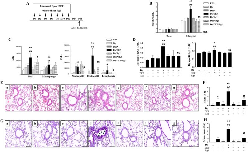
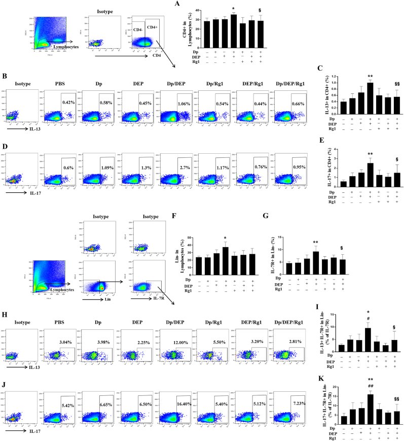
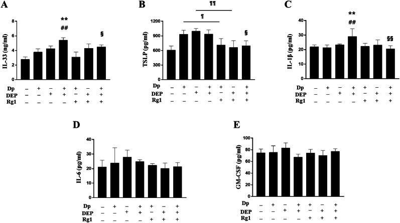
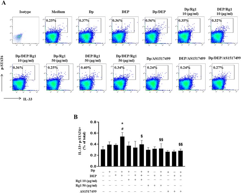
RESULTS Co-exposure to Dp and DEP significantly increased AHR, eosinophilic inflammation, Th2/Th17 responses, and ILC2/ILC3 populations. Ginsenoside Rg1 treatment markedly attenuated AHR, reduced eosinophils and serum Dp-specific IgG1, and decreased frequencies of IL-13+ ILC2s and IL-17+ ILC3s. IL-33 and IL-1β levels in lung tissue were also significantly reduced by Rg1. In MLE-12 cells, Rg1 suppressed IL-33 and phosphorylated STAT6 expression, mirroring the effects of a STAT6 inhibitor. Furthermore, Rg1 reduced IL-13 and IL-5 secretion from ILC2s co-cultured with MLE-12 cells.

CONCLUSION Ginsenoside Rg1 mitigates Dp/DEP-induced airway inflammation by downregulating Th2/Th17 and ILC2/ILC3 responses, potentially via the IL-33-STAT6 axis in airway epithelial cells.

© 2025 The Korean Society of Ginseng. Publishing services by Elsevier B.V.

Keywords

AnimalAsthmaDiesel exhaust particleDisease modelsEpithelial cellGinsenoside Rg1Innate lymphoid cell

Figures
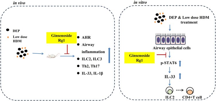
Fig. 1
Fig. 1 Effect of ginsenoside Rg1 on asthma features in mice exposed to Dp, DEP, or both (A) Experimental protocol. (B) Airway hyperresponsiveness in response to methacholine (Y axis: cmH2O·s/mL). (C) Inflammatory cell counts in bronchoalveolar lavage fluid, including total cells, macrophages, neutrophils, eosinophils, and lymphocytes. (D) Serum levels of Dp-specific IgG1, IgE (Y-axis: optical density, O.D.). (E–H) Histological analysis of lung tissues stained with Hematoxylin and Eosin (E) (original magnification × 200), inflammatory score (F), Periodic Acid–Schiff (G) (original magnification × 200) and mucus score (H). Black arrows indicate mucin secretion. Scale bars represent 100 μm. Experimental groups: a, PBS; b, Dp; c, DEP; d, Dp/DEP; e, Dp/Rg1; f, DEP/Rg1; g, Dp/DEP/Rg1. Five mice per group. Data are presented as mean ± standard deviation. Statistical significance: ∗P < 0.05, ∗∗P < 0.01 vs. Dp group; #P < 0.05, ##P < 0.01 vs. DEP group; §P < 0.05, §§P < 0.01 vs. Dp/DEP group. Statistical analysis was conducted using one-way ANOVA with Bonferroni's post hoc test. Abbreviations: Dp, Dermatophagoides pteronyssinus; DEP, diesel exhaust particles; Rg1, ginsenoside Rg1.
Fig. 2
Fig. 2 Effect of ginsenoside Rg1 on Th2/Th17 cells and ILC2s/ILC3s in lung cells of mice exposed to Dp, DEP, or both (A) Gating strategies and frequencies of CD4+ lymphocytes. (B, C) Gating plots and frequencies of IL-13+ in CD4+ lymphocytes (Th2 cells). (D, E) Gating plots and frequencies of IL-17+ in CD4+ lymphocytes (Th17 cells). The numbers state the percentage of cell subset per CD4+ lymphocytes. (F, G) Gating strategies and frequencies of IL-7Rα+ Lin-lymphocytes). (H, I) Gating plots and frequencies of IL-13+ ILC2s (IL-7Rα+ Lin-lymphocytes). (J, K) Gating plots and frequencies of IL-17+ ILC3s (IL-7Rα+ Lin-lymphocytes). The numbers state the percentage of cell subset per IL-7R + Lin-in lymphocytes. Five mice per group. Data are presented as mean ± standard deviation. Statistical significance: ∗P < 0.05, ∗∗P < 0.01 vs. Dp group; #P < 0.05, ##P < 0.01 vs. DEP group; §P < 0.05, §§P < 0.01 vs. Dp/DEP group. Statistical analysis was conducted using one-way ANOVA with Bonferroni's post hoc test. Abbreviations: Dp, Dermatophagoides pteronyssinus; DEP, diesel exhaust particles; Rg1, ginsenoside Rg1.
Fig. 3
Fig. 3 Effect of ginsenoside Rg1 on innate cytokine levels in lung homogenates of mice exposed to Dp, DEP, or both (A) IL-33. (B) TSLP. (C) IL-1β. (D) IL-6. (E) GM-CSF levels measured in crushed lung. Five mice per group. Data are presented as mean ± standard deviation. Statistical significance: ∗P < 0.05, ∗∗P < 0.01 vs. Dp group; #P < 0.05, ##P < 0.01 vs. DEP group; §P < 0.05, §§P < 0.01 vs. Dp/DEP group; P < 0.05, ¶¶P < 0.01 between two groups. Statistical analysis was conducted using one-way ANOVA with Bonferroni's post hoc test. Abbreviations: Dp, Dermatophagoides pteronyssinus; DEP, diesel exhaust particles; Rg1, ginsenoside Rg1; IL, interleukin; TSLP, thymic stromal lymphopoietin; GM-CSF, granulocyte-macrophage colony stimulating factor.
Fig. 4
Fig. 4 Evaluation of the effect of ginsenoside Rg1 or STAT6 inhibition on IL-33 and p-STAT6 expression in MLE-12 cells exposed to Dp, DEP, or both (A, B) Gating plots and frequencies of intracellular IL-33+ p-STAT6+ MLE12 cells expose to Dp (10 μg/ml), DEP (0.1 μg/ml) or both, with or without treatment of ginsenoside Rg1 (10 or 50 μg/ml) or the STAT6 inhibitor, AS1517499. Flow cytometry was used to assess intracellular marker expression. The numbers state the percentage of cell subset per total cells. Each group included four wells, and experiments were independently repeated three times with consistent results; representative data are shown. Data are presented as mean ± standard deviation. Statistical significance: ∗P < 0.05, ∗∗P < 0.01 vs. Dp group; #P < 0.05, ##P < 0.01 vs. DEP group; §P < 0.05, §§P < 0.01 vs. Dp/DEP group. Statistical analysis was conducted using one-way ANOVA with Bonferroni's post hoc test. Abbreviations: Dp, Dermatophagoides pteronyssinus; DEP, diesel exhaust particles; Rg1, ginsenoside Rg1; p-STAT6, phosphor-signal transducer and activator of transcription.
Fig. 5
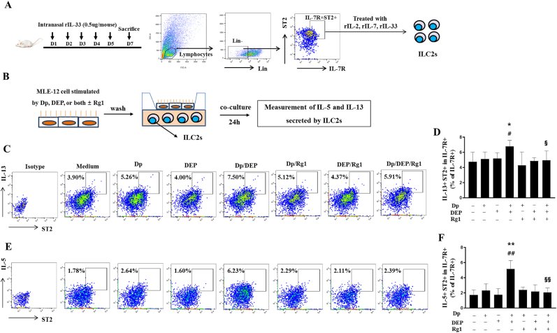
Fig. 5 Evaluation of the effect of ginsenoside Rg1 on IL-5 and IL-13 secretion from ILC2s co-cultured with MLE12 cells exposed to Dp, DEP, or both (A) Experimental procedure for mouse ILC2 isolation. (B) Schematic of the co-culture setup. (C, D) Gating plots and frequencies of ST2+IL-13+ producing cells from isolated and expanded ILC2s. (E. F) Gating plots and frequencies of ST2+IL-5+ producing cells from isolated and expanded ILC2s. The numbers state the percentage of cell subset per IL-7R + Lin-in lymphocytes. Each group included four wells, and experiments were independently repeated three times with consistent results; representative data are shown. Data are presented as mean ± standard deviation. Statistical significance: ∗P < 0.05, ∗∗P < 0.01 vs. Dp group; #P < 0.05, ##P < 0.01 vs. DEP group; §P < 0.05, §§P < 0.01 vs. Dp/DEP group. Statistical analysis was conducted using one-way ANOVA with Bonferroni's post hoc test. Abbreviations: Dp, Dermatophagoides pteronyssinus; DEP, diesel exhaust particles; Rg1, ginsenoside Rg1; ILC2, type 2 innate lymphoid cell.

그림 출처: PubMed Central 공개 논문 (PMC12805547)

Conflict of interest statement

The authors have no conflicting interests.

그 밖의 정보
출판 형태
Journal Article
저널
Journal of ginseng research · ISSN 1226-8453
전자 출판
2025-11-15
참고문헌
46편