Cigarette smoke extract enhances neutrophil elastase-induced IL-8 production via proteinase-activated receptor-2 upregulation in human bronchial epithelial cells.
Division of Pulmonary and Critical Care Medicine, Department of Internal Medicine, Seoul National University Hospital, Seoul, 03080, Korea.
Department of Internal Medicine, Seoul National University College of Medicine, Seoul, 03080, Korea.
Division of Pulmonary and Critical Care Medicine, Department of Internal Medicine, Seoul National University Hospital, Seoul, 03080, Korea. cgyoo@snu.ac.kr.
Department of Internal Medicine, Seoul National University College of Medicine, Seoul, 03080, Korea. cgyoo@snu.ac.kr.
Abstract
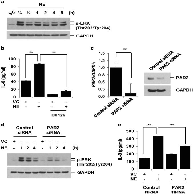
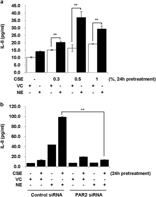
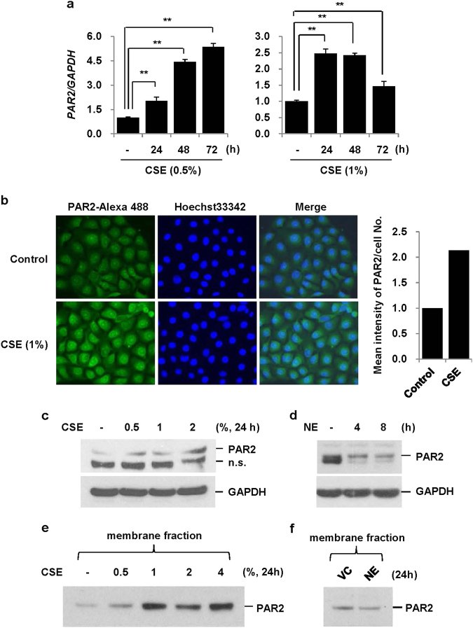
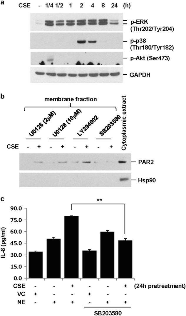
Although inflammation, oxidative stress, and protease-antiprotease imbalance have been referred to as a pathogenic triad in chronic obstructive pulmonary disease (COPD), little is known about how they interact. The objectives of this study were to elucidate the effect of cigarette smoke extract (CSE) on the neutrophil elastase (NE)-induced inflammatory response and its molecular mechanism in bronchial epithelial cells. We observed that NE activated extracellular signal-regulated kinase (ERK) and induced IL-8 production. Blocking ERK activation using a MEK inhibitor (U0126) suppressed NE-induced IL-8 secretion and knockdown of proteinase-activated receptor 2 (PAR2) using siRNAs inhibited both NE-induced ERK activation and subsequent IL-8 release, suggesting that NE-induced IL-8 production is dependent on PAR2-mediated ERK activation. Interestingly, pre-exposure to CSE markedly enhanced NE-induced IL-8 production. As PAR2 acts as a receptor for NE, we next investigated the effect of CSE on PAR2 expression as a molecular mechanism for the increased IL-8 production induced by NE in CSE exposed cells. CSE, but not NE, increased the expression of PAR2 mRNA and surface membrane protein. Inhibition of p38 MAPK reduced PAR2 expression induced by CSE while inhibition of the ERK and Akt pathway had no effect. Consequently, p38 inhibition significantly abrogated CSE-induced enhancement of IL-8 production in NE-treated cells. Of note, we observed increased PAR2 levels in lung homogenates and lung epithelial cells from CSE-treated mice and from both smokers and patients with COPD. Taken together, these results suggest that CSE upregulates PAR2 in normal human bronchial epithelial cells, thereby enhancing the inflammatory response to NE.
Figures
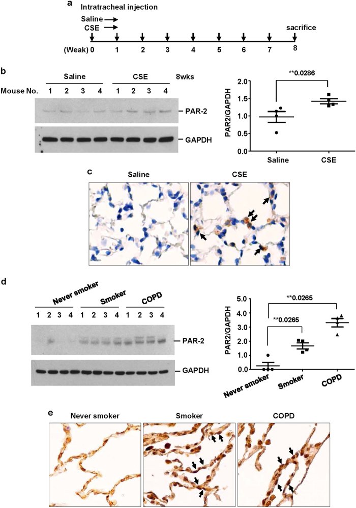
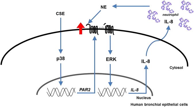
Fig. 1 NE increases IL-8 production via PAR2-mediated activation of ERK. a BEAS-2B cells were treated with vehicle control (VC) or NE (1 U/ml) for the indicated times. Total cellular extracts were subjected to western blot analysis for p-ERK and GAPDH. b Cells were pre-treated with an MEK inhibitor (U0126, 20 μM) for 1 h and then stimulated with VC or NE (1 U/ml) for 24 h. Levels of IL-8 in cell supernatants were measured by multiplex bead assay. Data represent the mean ± SD; **P < 0.05. c–e BEAS-2B cells were transiently transfected with control siRNAs or PAR2 siRNAs using a Neon electroporation kit. Forty-eight hours after transfection, cells were stimulated with VC or NE for 1, 2, 4 h (d) or 24 h (e). The expression of PAR2 was measured by quantitative real-time PCR. Data were normalized to the expression of GAPDH. Data represent the mean ± SD; **P < 0.05. Total cellular extracts were subjected to western blot analysis for PAR2, p-ERK and GAPDH. IL-8 concentrations in culture media were measured by multiplex bead assay. Data represent the mean ± SD; **P < 0.05Fig. 2 Pre-treatment with CSE enhances NE-induced IL-8 production via PAR2. a BEAS-2B cells were pre-treated with CSE (0.3, 0.5, and 1%) for 24 h and then stimulated with VC or NE (1 U/ml) for 24 h in the absence of CSE. b Cells were transiently transfected with control siRNAs or PAR2 siRNAs. Forty-eight hours after transfection, cells were pre-treated with CSE (1%) for 24 h and then stimulated with VC or NE (1 U/ml) for 24 h in the absence of CSE. IL-8 concentrations in cell supernatants were measured by multiplex bead assay. Data represent the mean ± SD; **P < 0.05Fig. 3 CSE, but not NE, upregulates PAR2 expression. a BEAS-2B cells were treated with CSE (0.5 and 1%) for the indicated times. The expression of PAR2 was measured by quantitative real-time PCR. Data were normalized to the expression of GAPDH. Data represent the mean ± SD; **P < 0.05. b Cells were stimulated with CSE (1%) for 24 h. The cells were fixed and permeabilized for 10 min. Immunofluorescent staining of PAR2 was performed using an anti-PAR2 antibody, followed by an Alexa Fluor 488 antibody. Cells were analyzed using an ECLIPSE TE300 (Nikon) fluorescence microscope. The mean intensity of PAR2 per cell number was determined using ImageJ. c, d BEAS-2B cells were treated with CSE (0.5, 1, and 2%) or NE (1 U/ml) for the indicated times. Total cell lysates were extracted and subjected to western blot analysis for PAR2 and GAPDH. e, f Cells were treated with CSE (0.5, 1, 2, and 4%) or NE (1 U/ml) for 24 h. The membrane fraction was isolated and then subjected to western blot analysis for PAR2 using an antibody to detect the N-terminal extracellular domain of PAR2. Results are representative of three independent experimentsFig. 4 CSE-induced upregulation of PAR2 is dependent on p38 activation. a BEAS-2B cells were treated with CSE (1%) for the indicated times. Total cellular extracts were subjected to western blot analysis for p-ERK, p-p38, p-Akt, and GAPDH. b Cells were pre-treated with an MEK inhibitor (U0126, 2 or 10 μM), PI3K/Akt inhibitor (LY294002, 10 μM), or p38 inhibitor (SB203580, 10 μM) for 2 h and then stimulated with CSE for 24 h in the presence of U0126, LY294002, or SB203580. Membrane and cytoplasmic proteins were extracted and subjected to western blot analysis for PAR2 and heat shock protein 90 (Hsp90). c BEAS-2B cells were pre-treated with SB203580 (10 μM) for 2 h and then stimulated with CSE (1%) in the presence or absence of SB203580 for 24 h. The cells were stimulated with VC or NE (1 U/ml) for 24 h in the absence of SB203580 and CSE. IL-8 concentrations in cell supernatants were measured by multiplex bead assay. Data represent the mean ± SD; **P < 0.05Fig. 5 Increased level of PAR2 found in lung homogenates and lung epithelial cells from CSE-treated mice and from smokers and COPD patients. a Experimental protocols for a murine model of emphysema. C57BL/6 mice were intratracheally instilled with vehicle or CSE as described in the materials and methods section. Mice (n = 4 per group) were sacrificed at week 8 after first instillation to isolate lungs. b Lung homogenates from mice were subjected to western blot analysis for PAR2 and GAPDH. Gel data were quantified using Scion image densitometry (right panel). Data represent the mean ± SE; **P < 0.05. c PAR2 immunohistochemistry in the lung tissues from mice. PAR2 was increased in lung epithelial cells of CSE-treated mice (arrows). d Total lung lysates obtained from normal (n = 4), smokers (n = 4), and COPD patients (n = 4) (c) were subjected to western blot analysis for PAR2 and GAPDH. Gel data were quantified (right panel). Data represent the mean ± SE; **P < 0.05. e PAR2 immunohistochemistry in the lung tissues from human patients. Arrows identify the epithelial cells. Original magnifications, ×200Fig. 6 Schematic diagram of the cross-talk between CSE and NE. CSE increases IL-8 production in lung epithelial cells. Released IL-8 recruits neutrophils into the lungs. Neutrophils secrete several proteases including NE. NE induces IL-8 production via the PAR2-mediated activation of ERK. CSE upregulates PAR2 through a p38-dependent pathway, which enhances NE-induced IL-8 production. A vicious cycle causing continued neutrophil accumulation and inflammation exists
그림 출처: PubMed Central 공개 논문 (PMC6035212)
Conflict of interest statement
The authors declare that they have no conflict of interest.
그 밖의 정보
출판 형태
Journal Article, Research Support, Non-U.S. Gov't
저널
Experimental & molecular medicine · eISSN 2092-6413