Department of Internal Medicine and Innovative Research Institute for Cell Therapy, Seoul National University Hospital, Seoul, Korea Biomedical Research Institute, Seoul National University Hospital, Seoul, Korea.
Department of Internal Medicine and Innovative Research Institute for Cell Therapy, Seoul National University Hospital, Seoul, Korea.
National Research Laboratory of Regenerative Sexual Medicine, Department of Urology, Inha University School of Medicine, Incheon, Korea.
Department of Internal Medicine and Innovative Research Institute for Cell Therapy, Seoul National University Hospital, Seoul, Korea Department of Molecular Medicine and Biopharmaceutical Sciences, Graduate School of Convergence Science and Technology, Seoul National University, Seoul, Korea hyosoo@snu.ac.kr.
Abstract
The importance of epigenetic regulation for maintenance of embryonic stem cell (ESC) pluripotency or for initiation of differentiation is widely accepted. However, the molecular mechanisms are poorly understood. We recently reported that a hypoxic microenvironment induces ESC differentiation. In the present study, we found that hypoxia-responsive histone deacetylase 6 (HDAC6) performs an essential signaling function for myogenic differentiation of ESCs. HDAC6 was downregulated in hypoxic ESCs or during differentiation. A knock-down of HDAC6 in ESCs resulted in induction of myogenic markers, including Pax7. Suppression of HDAC6 increased acetylation of core histones H3 and H4, leading to enhanced binding of RNA polymerase II to the Pax7 promoter. Transplantation of HDAC6 knock-down cells facilitated muscle regeneration in vivo. Importantly, the downregulation of HDAC6 by hypoxia was not mediated by HIF1α or HIF2α, master transcription regulators under hypoxia, but by induction of microRNA-26a that directly targeted the 3'-untranslated region (3'-UTR) of HDAC6. A point mutation of the microRNA-26a-binding sequence in the HDAC6 3'-UTR diminished the luciferase reporter activity. Taken together, these results suggest that environmental cues of differentiation modulate the epigenetic machinery and guide stem cells to commit to a specific lineage.
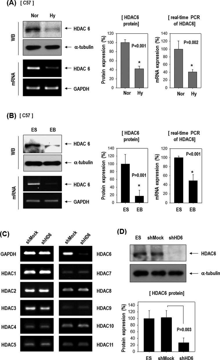
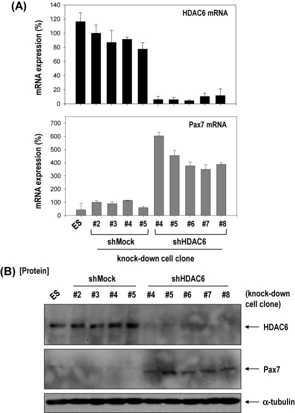
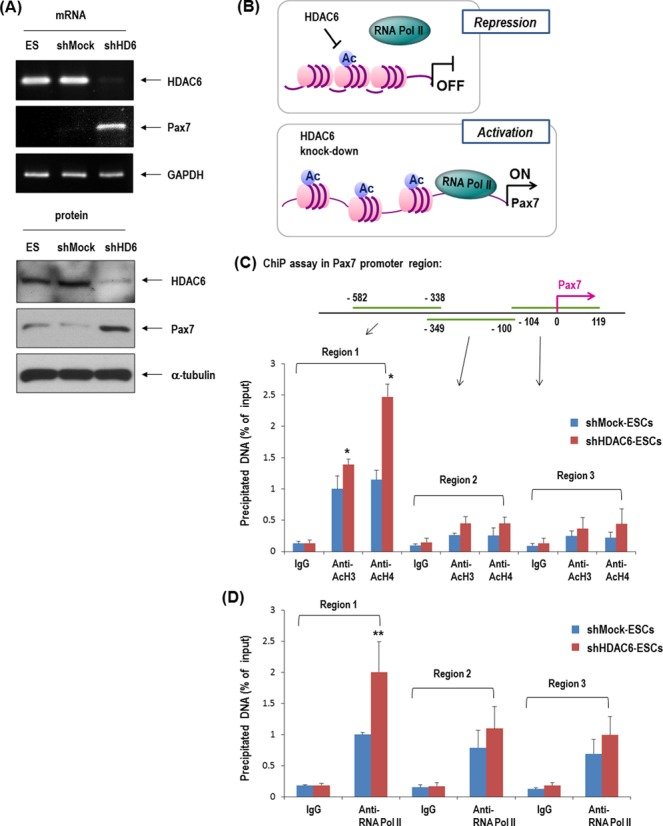
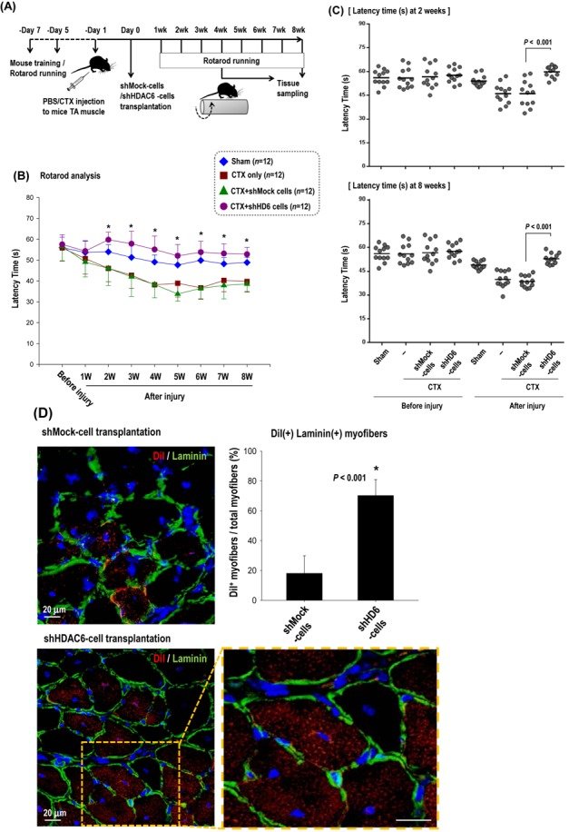
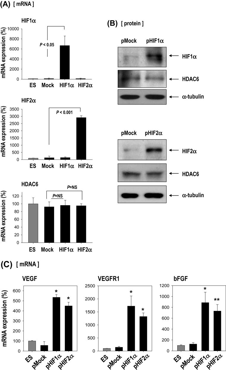
Figure 1.HDAC6 decreased in ESCs in response to hypoxia or during differentiation as EB formation and its knock-down stimulated genes of mesoderm- or myogenic-lineage. (A) HDAC6 protein and mRNA were reduced by 16 h hypoxia in C57 ESCs (n = 3). (B) HDAC6 protein and mRNA were reduced in EBs compared with ES (n = 3). (C and D) Generation of stable HDAC6 knock-down cells. HDAC6 mRNA (C) and protein (D) were markedly reduced by transfection of shHDAC6 (shHD6) compared with the non-target shRNA control (shMock) (n = 3). Especially, shHDAC6 transfection selectively suppressed HDAC6 among eleven HDAC family genes. (E) ESCs were cultured on a gelatin-coated plate in DMEM/10% FBS for 5 days, and real-time PCR was performed (n = 3). (F) Among mesoderm-lineage, myogenic-marker genes (Pax3, Pax7, MyoD) were markedly up-regulated in shHDAC6-cells upon differentiation, whereas vascular-marker genes (PECAM, SMA, VE-cadherin) were not (n = 4).Figure 2.Knock-down of HDAC6 activated differentiation of ECS toward myogenic-lineage. (A and B) Pax7, a marker of muscle progenitor, was remarkably increased at mRNA and protein levels in all five clones with HDAC6 knock-down C57 ESCs compared with all four control clones with shMock transfected. (C) Cells were plated onto a gelatin-coated dish in DMEM/10% FBS for 1 day, and further incubated up to 20 days under SkIM. (D) Pax7 and myogenic factors (Myf5, MyoD, Myogenin) increased after induction of differentiation to muscle in shMock-control cells, which was accentuated at greater degree in shHDAC6-cells (n = 3). (E) Immunofluorescence staining of shHDAC6-ESCs for Pax7 (red)/ nuclei (blue) on day10 or day 15 in SkIM. Positive control for Pax7 staining was myoblast cell line, C2C12 (×200).Figure 3.Knock-down of HDAC6 in ESCs induced epigenetic activation of Pax7 promoter. (A) Pax7 mRNA and protein remarkably increased in HDAC6 knock-down C57 ESCs. (B) Depiction of the regulation of histone acetylation and the recruitment of RNA polymerase II (Pol II) to the Pax7 promoter. The increase in the acetylation of H3 and H4 is a known indicator of active chromatin structure (40). (C and D) Quantification of ChIP assay with real-time PCR. Binding of acetylated histones H3 and H4 on Pax7 promoter was greater in HDAC6 knock-down than in control shMock. The binding of RNA polymerase II to the Pax7 promoter was also greater in shHDAC6 cells than in control ones (n = 4, *P < 0.001, **P < 0.05). (E) Western blot of acetylated histones H3 and H4 that were increased in shHDAC6 cells but were decreased by overexpression of HDAC6 in shHDAC6 cells. Quantification graph of western blot (n = 3).Figure 4.Knock-down of HDAC6 in ESCs facilitated muscle differentiation in vivo mouse model. (A–C) Rotarod test for mice with skeletal muscle injury. One day before transplantation, cardiotoxin (CTX) was injected into the tibialis anterior (TA) muscle (both legs) to induce muscle injury. We monitored the muscle power weekly for up to 8 weeks in four groups: sham control with PBS injection (sham, n = 12), injury control with CTX injection (CTX only, n = 12), injury with cell control (CTX+shMock, n = 12), injury with shHDAC6 cell transplantation (CTX+shHD6, n = 12). The measurement of the latency time for mice to fall off rotating rod at every week for up to 8 weeks (B) and at 2 weeks and 8 weeks (C) after cell transplantation were shown. (*P < 0.001, shMock-cells versus shHDAC6-cells). (D) Cross-section of TA muscle was immunostained for laminin α2 (green)/nuclei (blue) 4 weeks after transplantation of shHDAC6-ESCs that were pre-labeled with DiI. Magnification: ×400. Most of the red cells positive for DiI-fluorescence (red) were the regenerating myofibers with center nucleus. (E) Cross-sectional area of the regenerating myofibers in the injured TA muscles at 8 weeks after treatment with shMock-ESC or shHDAC6-ESC. Myofiber cross sectional area (CSA; μm2) was measured on H&E-stained cross-sections using the ImageJ software. Myofibers positive for DiI in the shHDAC6 group were bigger than those in the shMock group (top). CSA of the regenerating myofibers in the shHDAC6 group were significantly bigger than those in the shMock group (bottom, *P ≤ 0.001. Data are presented as mean ± SEM).Figure 5.HDAC6 reduction by hypoxia-responsive miR-26a in ESCs. (A–C) Effect of HIF1α or HIF2α over-expression on HDAC6 and other hypoxia-responsive control genes (VEGF, VEGFR1, bFGF). ESCs were transfected with pEGFP-HIF1α or pEGFP-HIF2α (n = 3). Real-time PCR and western blotting for HIF1α, HIF2α and HDAC6. Real-time PCR for control genes (VEGF, VEGFR1, bFGF) suggests that HIF plasmids functioned normally. (n = 3, *P < 0.001, **P < 0.05). (D) Sequence alignment of putative miR-22 and miR-26a targeting site within 3′UTR of HDAC6 (337 bp). (E–G) MicroRNA expression was measured by real-time PCR. (E) Induction of miR-22 and miR-26a by hypoxia in C57 or E14 ESCs (n = 4, *P < 0.001, **P < 0.01). (F) Suppression of HDAC6 only by miR-26a mimic but not by miR-22 in C57 ESCs (n = 3). (miR-NC: miRNA-negative control). (G) Suppression of HDAC6 only by pre-miR-26a plasmid (pmiR-26a) but not by pre-miR-22 plasmid (pmiR-22) in C57 cell (n = 3). (H) Western blot of HDAC6 after transfection of pmiR-22 and pmiR-26a. (I–K) miR-26a directly targeted 3′UTR of HDAC6 gene. (I) Schematic presentation of luciferase reporter constructs showing the predicted structures of each base-paired wild-type (WT) or mutant (mt1, mt2, mt3) 3′UTR of HDAC6. (J) Luciferase activity reflecting HDAC6 expression in C57 ESCs was suppressed by pmiR-26a but not by control pMock. Such inhibitory action of pmiR-26a was abrogated when target sites of 3′UTR were mutated (n = 3). (K) HDAC6 mRNA and protein expression were decreased by transfection of pre-miR-26a (pmiR-26a). Quantification graph of western blot (n = 4). (L) HDAC6 was not suppressed even under hypoxic conditions by transfection with AntagomiR (50 nM) against miR-26a (n = 3). (M) Model for HDAC6 suppression by hypoxia and myogenic differentiation of ESCs.