
PLoS One. 2014;9(8):e104354.
Circulating TNF receptors are significant prognostic biomarkers for idiopathic membranous nephropathy.
제1저자 이수미, 양승희 · 교신저자 이정표
소속 (8)
- Department of Internal Medicine, Seoul National University Hospital, Seoul, Korea; Department of Internal Medicine, Dong-A University, Busan, Korea.
- Department of Internal Medicine, Seoul National University Hospital, Seoul, Korea; Kidney Research Institute, Seoul National University, Seoul, Korea.
- Department of Internal Medicine, National Medical Center, Seoul, Korea.
- Department of Dental Hygiene, College of Health Science, Eulji University, Seongnam, Korea.
- Department of Internal Medicine, Seoul National University Hospital, Seoul, Korea; Department of Internal Medicine, Seoul National University Boramae Medical Center, Seoul, Korea.
- Department of Pathology, Seoul National University Bundang Hospital, Seongnam, Korea.
- Severance Biomedical Science Institute, Brain Korea 21 and Department of Internal Medicine, Yonsei University, Seoul, Korea.
- Department of Internal Medicine, Seoul National University Boramae Medical Center, Seoul, Korea.
Abstract
Idiopathic membranous nephropathy (iMN) is a common cause of nephrotic syndrome in adults. A biomarker to accurately indicate the severity of iMN and predict long-term prognosis is insufficient. Here, we evaluated the clinical significance of circulating tumor necrosis factor receptors (cTNFRs) as prognostic biomarkers of iMN with nephrotic syndrome. A total of 113 patients with biopsy-proven iMN and 43 healthy volunteers were enrolled in this study. Ninety patients with iMN had nephrotic range proteinuria. Levels of cTNFRs were measured by using serum samples collected at the time of initial diagnosis. Levels of cTNFRs were higher in the patients with nephrotic syndrome than in those with subnephrotic range proteinuria or in the healthy volunteers (P for trend <0.001). Estimated glomerular filtration rate and proteinuria tended to worsen as the cTNFRs levels increased. Having a cTNFR1 level within the highest tertile was a significant risk factor for renal progression after adjustment, in comparison with the other tertiles (hazard ratio [HR], 3.39; 95% confidence interval [95% CI], 1.48-7.78; P = 0.004). The cTNFR2 level within the highest tertile also significantly increased the risk of renal progression (HR, 3.29; 95% CI, 1.43-7.54; P = 0.005). Renal tubular TNFRs expression was associated with cTNFRs level. However, the cTNFRs levels were not associated with autoantibody against phospholipase A2 receptor reactivity/levels or treatment response. This study demonstrated that cTNFRs levels at the time of initial diagnosis could predict renal progression in patients with iMN.
Figures

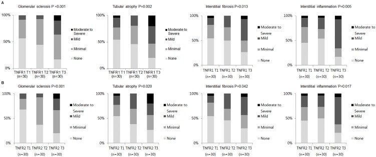


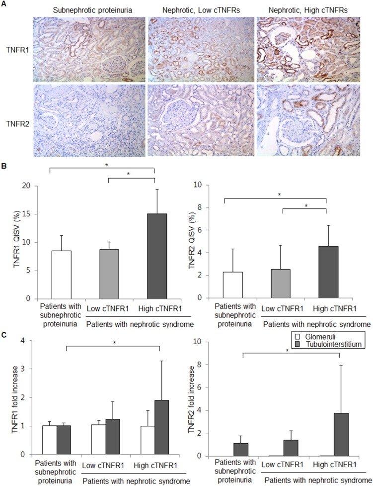
그림 출처: PubMed Central 공개 논문 (PMC4123977)
Conflict of interest statement
Competing Interests:The authors have declared that no competing interests exist.
그 밖의 정보
- 출판 형태
- Journal Article, Research Support, Non-U.S. Gov't
- 저널
- PloS one · eISSN 1932-6203
- 전자 출판
- 2014-08-06
- MeSH
- Adult · Aged · Biomarkers / blood · Female · Glomerular Filtration Rate* · Glomerulonephritis, Membranous* / blood / diagnosis / physiopathology · Humans · Male · Middle Aged · Nephrotic Syndrome* / blood / diagnosis / physiopathology · Prognosis · Receptors, Tumor Necrosis Factor, Type I / blood · Receptors, Tumor Necrosis Factor, Type II / blood
- Substances
- Biomarkers · Receptors, Tumor Necrosis Factor, Type I · Receptors, Tumor Necrosis Factor, Type II
- 참고문헌
- 44편