Cyclase-associated protein 1 is a binding partner of proprotein convertase subtilisin/kexin type-9 and is required for the degradation of low-density lipoprotein receptors by proprotein convertase subtilisin/kexin type-9.
National Leading Laboratory for Stem Cell Research, Seoul National University College of Medicine, 71, Daehak-Ro, Jongno-Gu, Seoul 03082, Korea.
Korea Research-Driven Hospital, Biomedical Research Institute, Seoul National University Hospital, 71, Daehak-ro, Jongro-gu, Seoul 03082, Korea.
Strategic Center of Cell & Bio Therapy, Seoul National University Hospital, 71, Daehak-ro, Jongro-gu, Seoul 03082, Korea.
Department of Cardiology, Asan Medical Center, University of Ulsan College of Medicine, 88, Olympic-ro 43-gil, Songpa-gu, Seoul 05505, Korea.
Department of Molecular Medicine and Biopharmaceutical Sciences, World Class University Program, Seoul National University, Seoul 03082, Korea.
Bio AI Research Center, Pharos I&BT Co., Ltd., Anyang-si, Gyeonggi-do 14059, Korea.
Division of Cardiology, Department of Internal Medicine, Severance Hospital, Yonsei University College of Medicine, 134 Shinchon-Dong, Seodaemun-Gu, Seoul 120752, Korea.
Department of Biochemistry and Molecular Biology, Brain Korea 21 PLUS Project for Medical Science, Yonsei University College of Medicine, Seoul 120752, Korea.
Department of Chemistry, Seoul National University, Seoul 120752, Korea.
Department of Convergence Medicine, University of Ulsan College of Medicine and Asan Medical Center, 88, Olympic-ro 43-gil, Songpa-gu, Seoul 05505, Korea.
Department of Internal Medicine, Seoul National University Hospital, 101, Daehak-Ro Jongno-Gu, Seoul 03080, Korea.
Cardiovascular Center & Department of Internal Medicine, Seoul National University Hospital, 101, Daehak-Ro Jongno-Gu, Seoul 03080, Korea.
Abstract
AIMS Proprotein convertase subtilisin/kexin type-9 (PCSK9), a molecular determinant of low-density lipoprotein (LDL) receptor (LDLR) fate, has emerged as a promising therapeutic target for atherosclerotic cardiovascular diseases. However, the precise mechanism by which PCSK9 regulates the internalization and lysosomal degradation of LDLR is unknown. Recently, we identified adenylyl cyclase-associated protein 1 (CAP1) as a receptor for human resistin whose globular C-terminus is structurally similar to the C-terminal cysteine-rich domain (CRD) of PCSK9. Herein, we investigated the role of CAP1 in PCSK9-mediated lysosomal degradation of LDLR and plasma LDL cholesterol (LDL-C) levels.
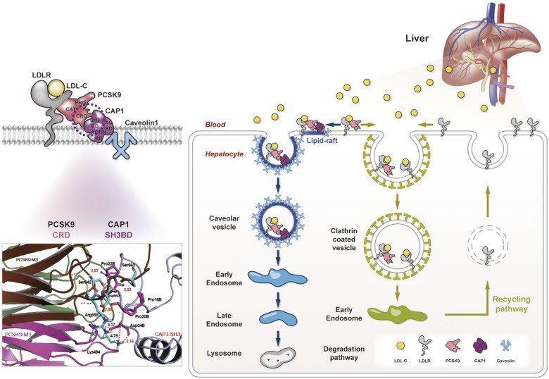
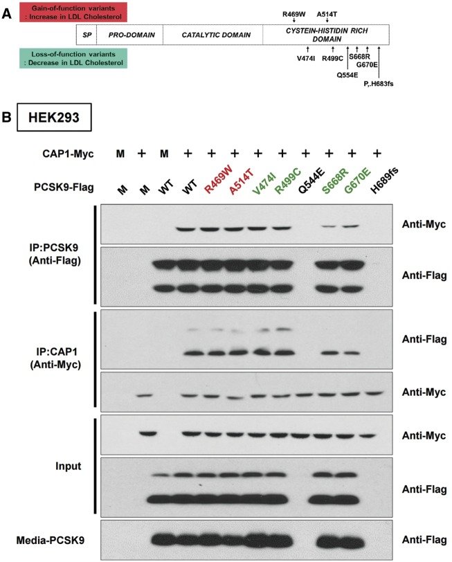
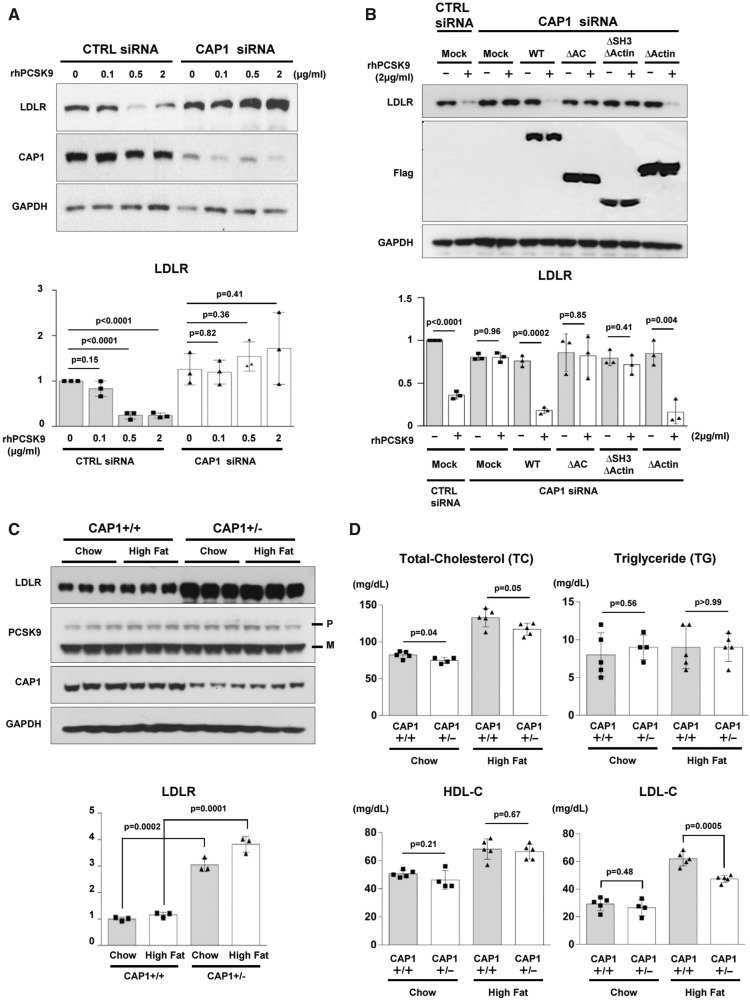
METHODS AND RESULTS The direct binding between PCSK9 and CAP1 was confirmed by immunoprecipitation assay, far-western blot, biomolecular fluorescence complementation, and surface plasmon resonance assay. Fine mapping revealed that the CRD of PCSK9 binds with the Src homology 3 binding domain (SH3BD) of CAP1. Two loss-of-function polymorphisms found in human PCSK9 (S668R and G670E in CRD) were attributed to a defective interaction with CAP1. siRNA against CAP1 reduced the PCSK9-mediated degradation of LDLR in vitro. We generated CAP1 knock-out mice and found that the viable heterozygous CAP1 knock-out mice had higher protein levels of LDLR and lower LDL-C levels in the liver and plasma, respectively, than the control mice. Mechanistic analysis revealed that PCSK9-induced endocytosis and lysosomal degradation of LDLR were mediated by caveolin but not by clathrin, and they were dependent on binding between CAP1 and caveolin-1.
CONCLUSION We identified CAP1 as a new binding partner of PCSK9 and a key mediator of caveolae-dependent endocytosis and lysosomal degradation of LDLR.
CaveolaeCyclase-associated protein 1EndocytosisLDL cholesterolLow-density lipoprotein receptorProprotein convertase subtilisin/kexin type-9
Figures
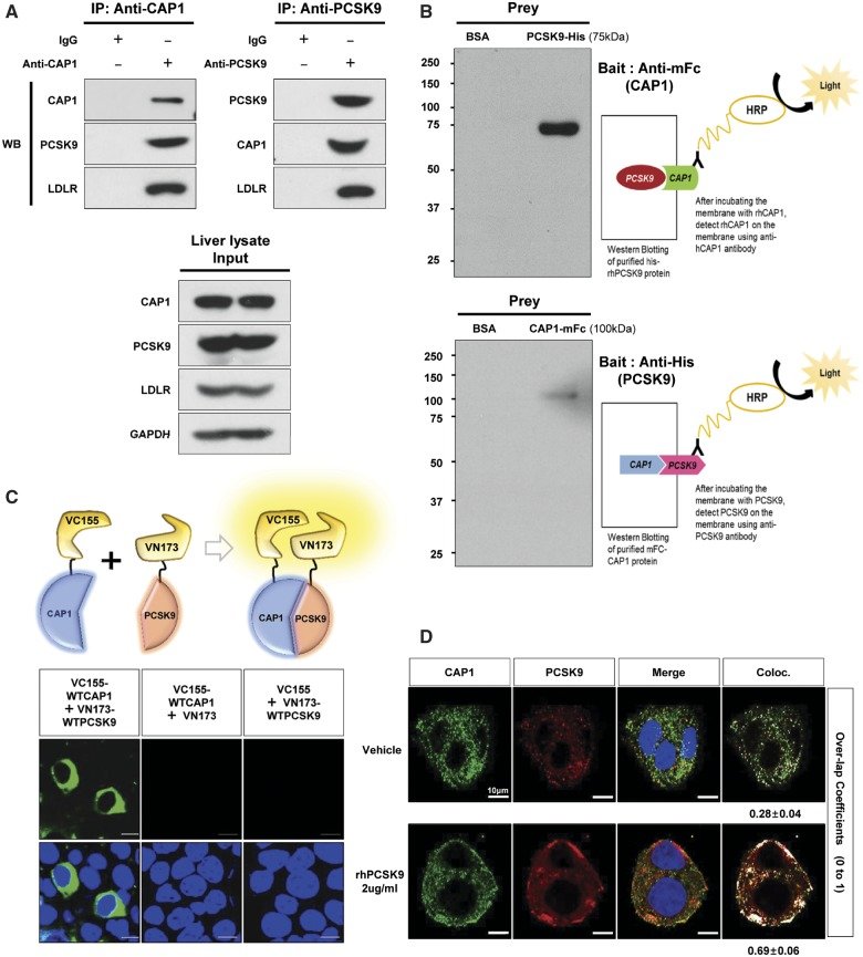
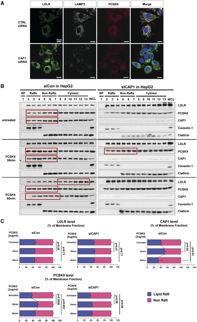
Figure 1CAP1 directly interacts with PCSK9. (A) Immunoprecipitation of endogenous CAP1 or PCSK9 from liver lysates of C57/BL6 wild-type mice. (B) Far-western blot analysis of mFc-hCAP1 and hPCSK9-His. (C) Bimolecular fluorescence complementation assay visualizing the interaction between hCAP1 and hPCSK9 in living cells. Green fluorescence was observed when CAP1 and PCSK9 fused to each fragment (pVC155 and pVN173) and were both expressed (left panel). However, when human CAP1 or PCSK9 was expressed alone, no fluorescence was detected (middle and right panel, respectively). (D) Colocalization of CAP1 and PCSK9 as determined by immunofluorescent staining of HepG2 cells exogenously treated with recombinant hPCSK9. Overlap coefficients: mean ± standard deviation. (E) Direct binding assay between rhPCSK9 and CAP1 using surface plasmon resonance. The equilibrium dissociation constant was 1.01 µM. (F) Coimmunoprecipitation of hPCSK9-Flag and wild-type CAP1 or each CAP1 mutant in HEK 293 cells. Pro-PCSK9 (P) and mature PCSK9 (M) are indicated with bold line. (G) Coimmunoprecipitation of hPCSK9-Flag and the SH3BD of CAP1 in HEK 293 cells showing that the SH3BD is sufficient for binding to CAP1. (H) Coimmunoprecipitation of wild-type hCAP1 with wild-type PCSK9 or the CRD deletion PCSK9 mutant in HEK 293 cells showing that PCSK9 binds CAP1 via the CRD.Figure 2Human PCSK9 loss-of-function mutants have weaker interactions with CAP1 than the wild type. (A) The single-nucleotide variants of PCSK9-CRD reported to be associated with plasma LDL cholesterol levels in humans. (B) The loss-of-function PCSK9 mutants S668R and G679E lose their binding affinity to CAP1. CAP1-Myc and either wild-type PCSK9-Flag or each point mutant of PCSK9-Flag were overexpressed in HEK293 cells. Immunoprecipitation with Flag and immunoblotting with Myc (upper two figures of immunoblot), or immunoprecipitation with Myc and immunoblotting with Flag (immunoblot on the third and fourth lines). The level of PCSK9 in the culture media (the lowest line).Figure 3CAP1 is required for PCSK9-induced LDLR degradation. (A) LDLR degradation induced by PCSK9 was attenuated by CAP1 siRNA (upper panel). The representative figure of three independent experiments. Quantification of the expression levels of LDLR (bottom panel). (B) PCSK9-induced LDLR degradation was rescued by wild-type CAP1 and the ▵ACBD mutant (upper panel). Quantification of the expression levels of LDLR (bottom panel). (C) The protein expression levels of LDLR were significantly greater in the CAP1+/− mice than those in the CAP1+/+ mice, regardless of the diet. Pro-PCSK9 (P) and mature PCSK9 (M) are indicated with bold line. Quantification of the expression levels of LDLR/GAPDH in CAP1+/− vs. CAP1+/+ mice (bottom panel). (D) Enzymatic measurement of the plasma cholesterol levels in CAP1+/− and CAP1+/+ mice fed either high fat- or chow diet for 16 weeks (n = 6 per group). (E) Cholesterol levels in FPLC fractions from pooled plasma samples (n = 5 per group) in CAP+/− (red) and CAP1+/+ (blue) mice fed high-fat diet. (F–H) Overexpression of PCSK9 in both CAP1+/− and CAP1+/+ mice using adeno-associated virus. (F) Immunoblot analysis of PCSK9-induced LDLR degradation in CAP1+/− and CAP1+/+ mice liver. Pro-PCSK9 (P) and mature PCSK9 (M) are indicated with bold line. The representative figure of three independent experiments. Quantification of the expression levels of LDLR (bottom left), PCSK9 (bottom middle), and CAP1 (bottom right) (n = 9). (G) Plasma levels of hPCSK9 were measured by ELISA (n = 5 per group). (H) Enzymatic analysis of plasma cholesterol levels in CAP1+/− and CAP1+/+ mice with or without PCSK9 overexpression (n = 5 per group).Figure 4CAP1 mediates the caveolin-dependent endocytosis and lysosomal degradation of LDLR. (A) Immunofluorescent staining of HepG2 cells with LDLR (green), PCSK9 (red), and the lysosomal marker LAMP2 (white) 240 min after treatment with recombinant hPCSK9. (B) Membrane fractionation and immunoblot analysis demonstrating the cellular distribution of LDLR, PCSK9 and CAP1 after PCSK9 treatment in HepG2 cells treated with either a control or CAP1 siRNA. The representative figure of three independent experiments. (C) Quantification of the immunoblot. (D and E) Co-localization of endosomal marker EEA1 with clathrin. . (D) and caveolin-1 (E) in HepG2 cells treated with recombinant rhPCSK9 for 30 min. Overlap coefficients: mean ± standard deviation. (F and G) Immunoblot analysis of LDLR expression demonstrating that PCSK9-induced LDLR degradation was attenuated by caveolin siRNA (F) in HepG2 cells but was not affected by clathrin siRNA (G). Quantification of the expression levels of LDLR (bottom panel, n = 3). (H) Immunofluorescent staining of HepG2 cells with LDLR (green) and the lysosomal marker LAMP2 (red) 240 min after hPCSK9 treatment. HepG2 cells were pre-treated with either the control, CAP1, caveolin1, or clathrin siRNA. (I) Immunofluorescent staining of HepG2 cells with EGF (red) and albumin (green). Bar indicates 10 μm. The representative figure of three independent experiments, and the dots in a cell were counted from ten random fields of each independent experiment (bottom panel). (J) LDLR degradation by the overexpression of PCSK9 was attenuated by CAP1 siRNA and caveolin1 siRNA but not clathrin siRNA. Bar graphs showing the LDLR and PCSK9 level in the lysate. The representative figure of three independent experiments. (K) Transmission electron microscopic images of HepG2 cells without treatment (upper left) or with pretreatment with either a control siRNA (lower left) or CAP1 siRNA (lower right) and exposed to PCSK9 for 15 min. Numerous caveolae (yellow circle), caveolin-vesicles (yellow arrows), and clathrin-coated vesicles (red arrows) are observed in the control siRNA-PCSK9-treated cells, while only clathrin-coated vesicles are observed in the CAP1 siRNA-PCSK9-treated cells. Bar indicates 0.5 μm; ER, endoplasmic reticulum; MT, mitochondrion; N, nucleus. (L) hPCSK9-Flag and either wild-type CAP1-Myc or its mutants were overexpressed in HEK 293 cells. Immunoprecipitation with anti-Flag beads and immunoblotting with Myc or caveolin-1. (M) LDLR localizes to the recycling endosomes in PCSK9-deficient cells but not much in clathrin-deficient cells. Immunofluorescent staining of HepG2 cells ‘at 4 h after LDL-C treatment’ with Rab11 (red), LDLR (green), and DAPI (blue). Colocalization of Rab11 and LDLR was measured and expressed as percent colocalization (bottom graph). (N) Schematic model in which LDLR undergoes caveolae-dependent endocytosis and lysosomal degradation induced by PCSK9. CAP1 mediates this process by interfacing PCSK9 with caveolin through the SH3BD and ACBD.Take home figureCAP1 is a new binding partner of PCSK9 and mediates caveolae-dependent endocytosis and lysosomal degradation of LDLR.