Division of Pulmonary and Critical Care Medicine, Department of Internal Medicine, Seoul National University Hospital, Seoul 03080, Korea.
Department of Internal Medicine, Seoul National University College of Medicine, Seoul 03080, Korea.
Abstract
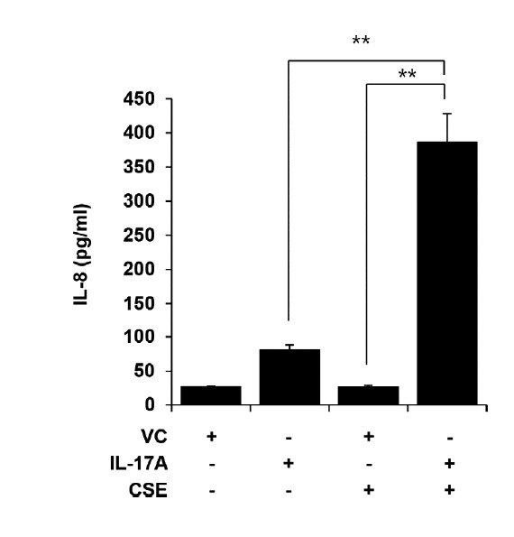
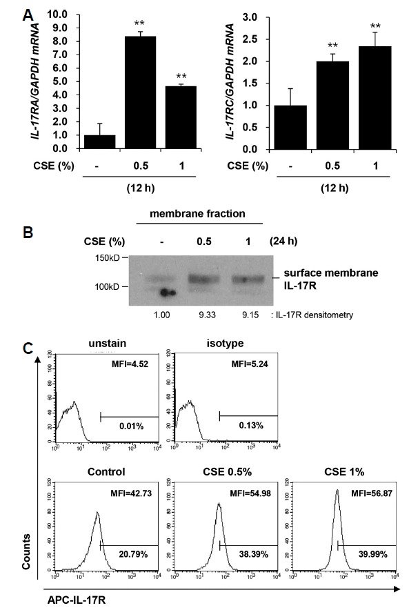
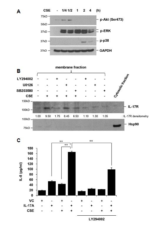
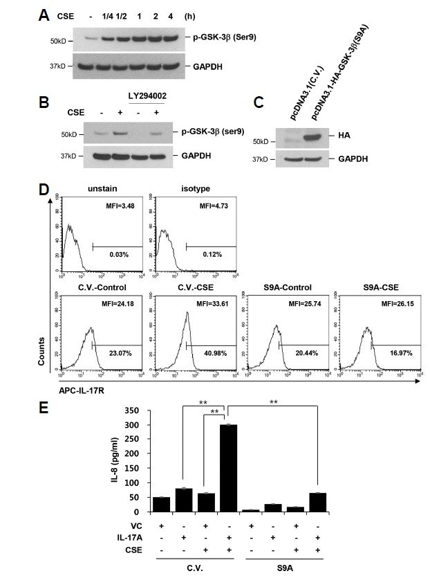
Interleukin-17A (IL-17A) is a pro-inflammatory cytokine mainly derived from T helper 17 cells and is known to be involved in the pathogenesis of chronic obstructive pulmonary disease (COPD). Cigarette smoke (CS) has been considered as a primary risk factor of COPD. However, the interaction between CS and IL-17A and the underlying molecular mechanisms have not been clarified. In the current study, we investigated the effects of cigarette smoke extract (CSE) on IL-17A-induced IL-8 production in human bronchial epithelial cells, and sought to identify the underlying molecular mechanisms. IL-8 production was significantly enhanced following treatment with both IL-17A and CSE, while treatment with either IL-17A or CSE alone caused only a slight increase in IL-8 production. CSE increased the transcription of IL-17RA/RC and surface membrane expression of IL-17R, which was suppressed by an inhibitor of the phosphoinositide 3-kinase (PI3K)/Akt pathway (LY294002). CSE caused inactivation of glycogen synthase kinase-3β (GSK-3β) via the PI3K/Akt pathway. Blockade of GSK-3β inactivation by overexpression of constitutively active GSK-3β (S9A) completely suppressed the CSE-induced up-regulation of IL-17R expression and the CSE-induced enhancement of IL-8 secretion. In conclusion, inactivation of GSK-3β via the PI3K/Akt pathway mediates CSE-induced up-regulation of IL-17R, which contributes to the enhancement of IL-17A-induced IL-8 production.
Keywords
AktGSK-3βIL-17AIL-17RIL-8cigarette smoke extract
Figures
Fig. 1 CSE enhances IL-17A-induced IL-8 production BEAS-2B cells were treated with CSE (1%) in the presence or absence of IL-17A (100 ng/ml) for 24 h. IL-8 concentration in cell culture media was measured using a multiplex bead assay. Data represent mean ± SD of triplicates. **P < 0.05. Results are representative of three independent experiments.Fig. 2 CSE up-regulates IL-17R expression BEAS-2B cells were treated with CSE (0.5, 1%) for 12 h (A) or 24 h (B, C). (A) Total RNA was isolated and quantitative real-time PCR for IL-17RA, IL-17RC, and GAPDH was performed. Data represent mean ± SD of triplicates. **P < 0.05 versus control. (B) Membrane fraction was isolated and then subjected to western blot analysis for IL-17R to detect the N-terminal extracellular domain of IL-17RA/RC. (C) Cell surface expression of IL-17R was determined by flow cytometry. Results are shown as mean fluorescence intensity (MFI) histograms. Results are representative of three independent experiments.Fig. 3 Enhanced IL-17A-induced IL-8 production by CSE is dependent on PI3K/Akt activation (A) BEAS-2B cells were treated with CSE for the indicated times. Total cellular extracts were subjected to Western blot analysis for p-Akt (Ser473), p-ERK (Thr202/Tyr204), p-p38 (Thr180/Tyr182), and GAPDH. (B) Cells were pretreated with a MEK1/2 inhibitor (U0126, 2 μM), PI3K/Akt inhibitor (LY294002, 10 μM), or p38 inhibitor (SB203580, 10 μM) for 2 h then stimulated with CSE for 24 h in the presence of U0126, LY294002, or SB203580. Membrane proteins were extracted and subjected to Western blot analysis for IL-17R. Cytosolic proteins were subjected to western blot analysis for heat shock protein 90 (Hsp90). (C) BEAS-2B cells were pretreated with LY294002 (10 μM) for 1 h then stimulated with CSE and IL-17A in the presence or absence of LY294002 for 24 h. IL-8 concentration in culture media was determined using a multiplex bead assay. Data represent mean ± SD of triplicates. **P < 0.05. Results are representative of three independent experiments.Fig. 4 Increased IL-17R through PI3K/Akt activation is mediated by GSK-3β inactivation (A) BEAS-2B cells were treated with CSE for the indicated times. Expression of p-GSK-3β (Ser9) was assessed by Western blot analysis. (B) Cells were pretreated with PI3K/Akt inhibitor (LY294002, 10 μM) for 1 h then stimulated with CSE in the presence or absence of LY294002 for 1 h. Total cellular extracts were subjected to Western blot analysis for p-GSK-3β (Ser9) and GAPDH. (C–E) Cells were transiently transfected with an HA-tagged GSK-3β (S9A) expression vector or a control vector (C.V., pcDNA3.1). After 48 h, cells were treated with CSE for 24 h. Total cellular extracts were subjected to western blot analysis for HA and GAPDH (C). Cell surface expression of IL-17R was determined by flow cytometry (D). IL-8 concentration in culture media was determined using a multiplex bead assay (E). Data represent mean ± SD of triplicates. **P < 0.05. Results are representative of three independent experiments.