Division of Pulmonary and Critical Care Medicine, Department of Internal Medicine, Seoul National University Hospital, Seoul 03080, Korea.
Department of Internal Medicine, Seoul National University College of Medicine, Seoul 03080, Korea.
Abstract
Adiponectin, a hormone produced by adipose tissue, is very abundant in plasma, and its anti- and pro-inflammatory effects are reported. However, the mechanisms of these pro- and anti-inflammatory effects are not fully defined. Herein, we evaluated the dual inflammatory response mechanism of adiponectin in macrophages. Short-term globular adiponectin (gAd) treatment induced IκBα degradation, NF-κB nuclear translocation, and TNF-α production in RAW 264.7 cells. Polymyxin B pretreatment did not block gAd-induced IκBα degradation, and heated gAd was unable to degrade IκBα, suggesting that the effects of gAd were not due to endotoxin contamination. gAd activated IKK and Akt, and inhibition of either IKK or Akt by dominant-negative IKKβ (DN-IKKβ) or DN-Akt overexpression blocked gAd-induced IκBα degradation, suggesting that short-term incubation with gAd mediates inflammatory responses by activating the IκB/NF-κB and PI3K/Akt pathways. Contrastingly, long-term stimulation with gAd induced, upon subsequent stimulation, tolerance to gAd, lipopolysaccharide, and CpG-oligodeoxynucleotide, which is associated with gAd-induced downregulation of IL-receptor-associated kinase-1 (IRAK-1) due to IRAK-1 transcriptional repression. Conclusively, our findings demonstrate that the pro- and anti-inflammatory responses to gAd in innate immune cells are time-dependent, and mediated by the activation of the IκB/NF-κB pathway, and IRAK-1 downregulation, respectively.
Keywords
IRAK-1NF-κBglobular adiponection
Figures
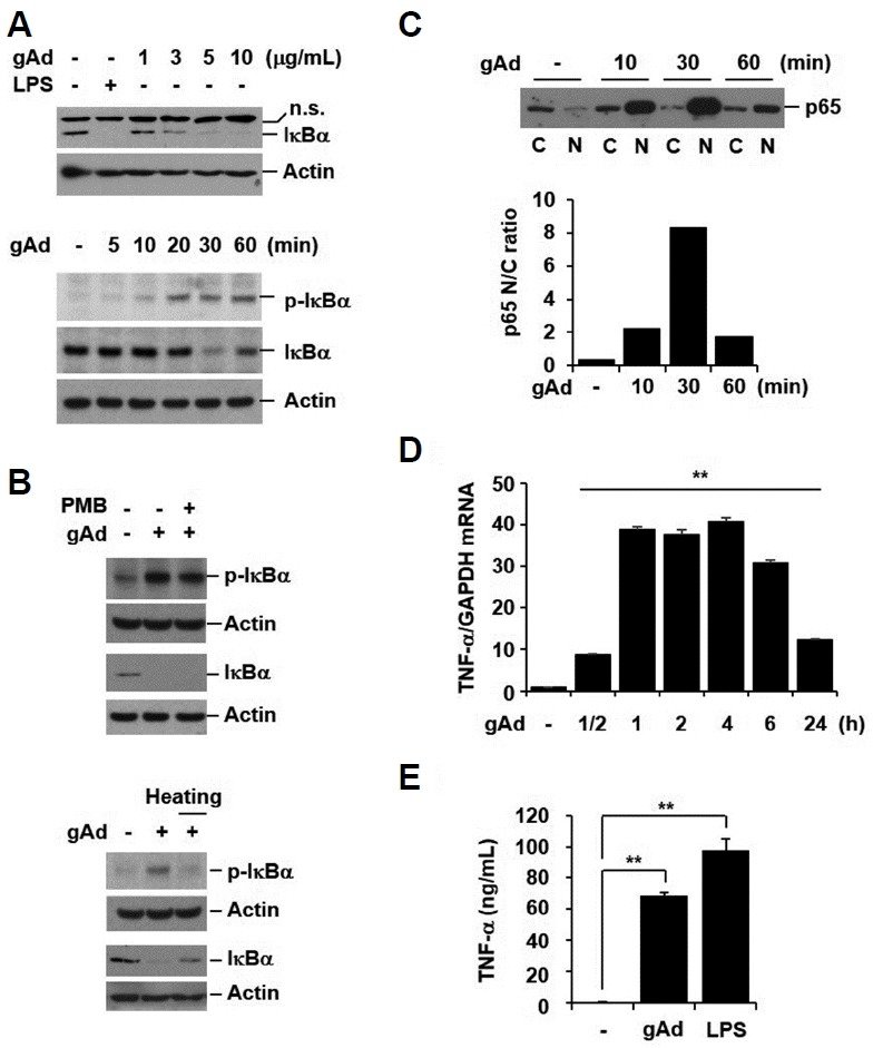
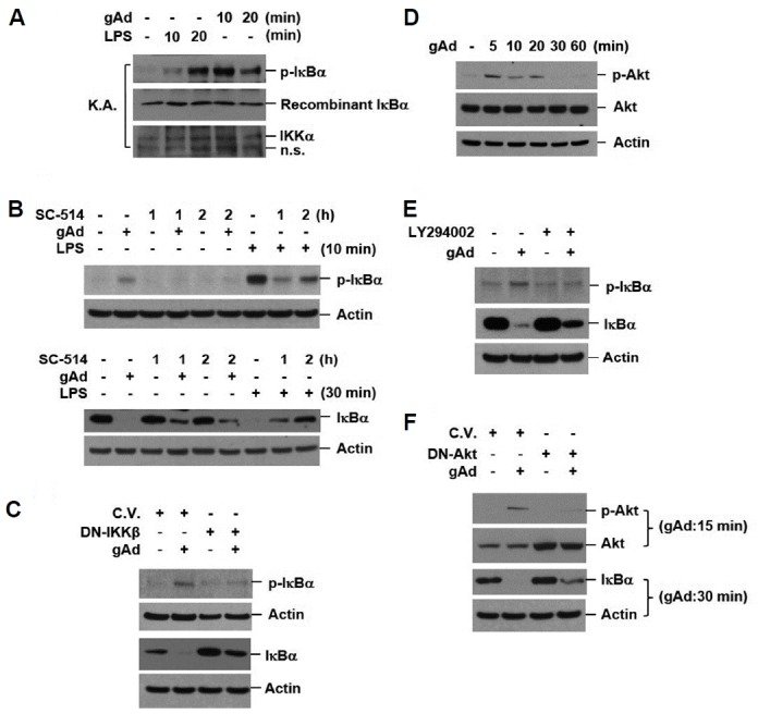
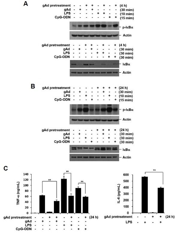
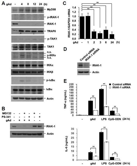
Fig. 1 gAd activates NF-κB and induces pro-inflammatory cytokine secretion (A) RAW 264.7 cells were incubated with gAd (1, 3, 5, 10 μg/ml) and LPS (1 μg/ml) for 30 min. Cells were stimulated with gAd (5 μg/ml) for the indicated times. (B) RAW 264.7 cells were pretreated with polymyxin B (PMB, 10 μg/ml) for 1 h, and then stimulated with gAd (5 μg/ml) for 30 min in the presence or absence of PMB. Cells were treated with gAd and heated gAd (5 μg/ml) for 30 min. Total cellular extracts were subjected to Western blot analysis for p-IκBα, IκBα and actin. (C) Cells were stimulated with gAd (5 μg/ml) for the indicated times. Cytoplasmic (C) and nuclear (N) proteins were extracted. Equal amounts of protein were subjected to Western blot analysis for p65. Ratio of nuclear to cytoplasmic (N/C) p65 densitometry values (lower panel of (C)). (D) RAW 264.7 cells were stimulated with gAd (5 μg/ml) for the indicated times. Total RNA was isolated and quantitative real-time PCR for TNF-α and GAPDH was performed. Data represent the mean ± SD of triplicates. **P < 0.05 (E) Cells were treated with gAd (5 μg/ml) and LPS (1 μg/ml) for 24 h. TNF-α concentration in culture medium was measured by multiplex bead assay. Data represent the mean ± SD of triplicates. **P < 0.05 Results are representative of three separate experiments. n.s., non-specific.Fig. 2 gAd-induced IκBα degradation is mediated by IKK and Akt activation (A) RAW 264.7 cells were treated with gAd (5 μg/ml) and LPS (1 μg/ml) for the indicated times. The IKK complex was immunoprecipitated using anti-IKKα antibodies. The kinase activity of IKK was assayed as described in the materials and methods section. (B) RAW 264.7 cells were preincubated with SC-514 (an IKKβ specific inhibitor, 100 μM) and then, stimulated with gAd (5 μg/ml) for 30 min and LPS (1 μg/ml) for 10 or 30 min in the presence or absence of SC-514. (C) Cells were transiently transfected with DN-IKKβ (K44A) and control plasmid vector (C.V.). At 48 h after transfection, cells were incubated with gAd (5 μg/ml) for 30 min. (D) Cells were treated with gAd (5 μg/ml) for the indicated times. (E) RAW 264.7 cells were pretreated with LY294002 (40 μM) for 2 h and then stimulated with gAd (5 μg/ml) for 30 min. (F) Cells were transfected with DN-Akt and control plasmid vector. At 48 h after transfection, cells were incubated with gAd (5 μg/ml) for 15 or 30 min. Total cellular extracts were subjected to Western blot analysis for p-IκBα, IKKα, IκBα, p-Akt, Akt, and actin. Results are representative of three separate experiments. K.A., kinase activity assay.Fig. 3 gAd pretreatment suppresses inflammatory response to gAd, LPS, and CpG-ODN (A, B) RAW 264.7 cells were preincubated with gAd (5 μg/ml) for 4 h or 24 h, and then, washed several times with fresh medium. The cells were stimulated with gAd (5 μg/ml), LPS (1 μg/ml), and CpG-ODN (1 μM) for the indicated times. Total cellular extracts were subjected to Western blot analysis for p-IκBα, IκBα, and actin. (C) After preincubation with gAd for 24 h, RAW 264.7 cells were washed, and then stimulated with gAd, LPS and CpG-ODN for 20 h. TNF-α and IL-6 concentrations in the culture supernatant were measured by multiplex bead assay. Data represent the mean ± SD of triplicates. **P < 0.05 Results are representative of three separate experiments.Fig. 4 gAd downregulates IRAK-1 mRNA and protein expression (A) RAW 264.7 cells were incubated with gAd (5 μg/ml) for the indicated times. (B) Cells were pretreated with MG132 (10 μM) and PS-341 (200 nM) for 1 h, and then stimulated with gAd (5 μg/ml) for 4 h in the presence or absence of MG132 and PS-341. Total cellular extracts were subjected to Western blot analysis for MyD88, p-IRAK-1, IRAK-1, TRAF6, p-TAK1, TAK1, p-IKKα/β, IKKα, IKKβ, p-IκBα, IκBα, and actin. (C) Cells were treated with gAd (5 μg/ml) for the indicated times. Quantitative real-time PCR for IRAK-1 and GAPDH was performed. Data represent the mean ± SD of triplicates. **P < 0.05 (D-E) RAW 264.7 cells were transiently transfected with control or IRAK-1 siRNAs. At 48 h after transfection, cells were treated with gAd (5 μg/ml), LPS (1 μg/ml), or CpG-ODN (1 μM) for 24 h. Total cellular extracts were subjected to Western blot analysis for IRAK-1 and Actin. TNF-α and IL-6 concentrations in culture medium were measured by multiplex bead assay. Data represent the mean ± SD of triplicates. **P < 0.05 Results are representative of three separate experiments.