Division of Pulmonary and Critical Care Medicine, Department of Internal Medicine, Seoul National University Hospital, Seoul, Korea.
Department of Internal Medicine, Seoul National University College of Medicine, Seoul, Korea.
Abstract
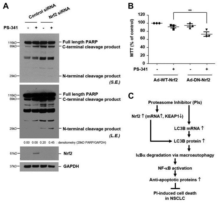
IκB, a cytoplasmic inhibitor of nuclear factor-κB (NF-κB), is reportedly degraded via the proteasome. However, we recently found that long-term incubation with proteasome inhibitors (PIs) such as PS-341 or MG132 induces IκBα degradation via an alternative pathway, lysosome, which results in NF-κB activation and confers resistance to PI-induced lung cancer cell death. To enhance the anti-cancer efficacy of PIs, elucidation of the regulatory mechanism of PI-induced IκBα degradation is necessary. Here, we demonstrated that PI upregulates nuclear factor (erythroid-derived 2)-like 2 (Nrf2) via both de novo protein synthesis and Kelch-like ECH-associated protein 1 (KEAP1) degradation, which is responsible for IκBα degradation via macroautophagy activation. PIs increased the protein level of light chain 3B (LC3B, macroautophagy marker), but not lysosome-associated membrane protein 2a (Lamp2a, the receptor for chaperone-mediated autophagy) in NCI-H157 and A549 lung cancer cells. Pretreatment with macroautophagy inhibitor or knock-down of LC3B blocked PI-induced IκBα degradation. PIs up-regulated Nrf2 by increasing its transcription and mediating degradation of KEAP1 (cytoplasmic inhibitor of Nrf2). Overexpression of dominant-negative Nrf2, which lacks an N-terminal transactivating domain, or knock-down of Nrf2 suppressed PI-induced LC3B protein expression and subsequent IκBα degradation. Thus, blocking of the Nrf2 pathway enhanced PI-induced cell death. These findings suggest that Nrf2-driven induction of LC3B plays an essential role in PI-induced activation of the IκB/NF-κB pathway, which attenuates the anti-tumor efficacy of PIs.
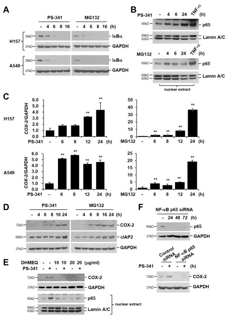
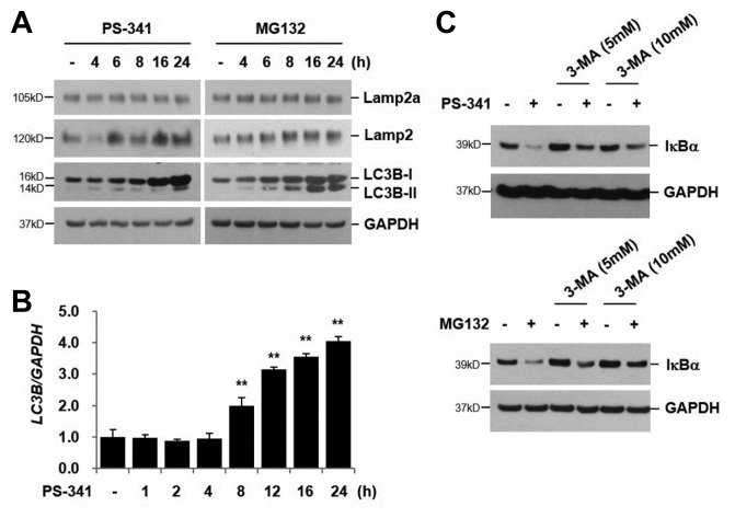
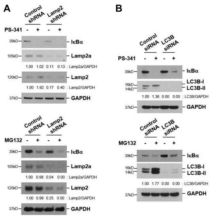
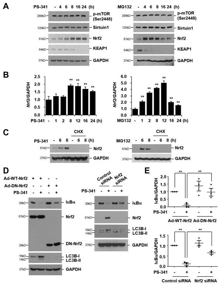
Fig. 1 PI activates IκB/NF-κB pathway (A) NCI-H157 and A549 cells were treated with PS-341 (50 nM) or MG132 (20 μM) for the indicated times. Total cellular extracts were subjected to Western blot analysis for IκBα and GAPDH. (B) NCI-H157 cells were treated with PS-341 or MG132 for the indicated times. Nuclear proteins were extracted and subjected to Western blot analysis for p65 and Lamin A/C. TNF-α (10 ng/mL, 60 min) was used as a positive control. (C) Both cell lines were stimulated with PS-341 or MG132 for 0, 6, 8, 12, or 24 h. Total RNA was isolated and quantitative real-time PCR for COX-2 and GAPDH was performed. Data represent the mean ± SD of triplicate experiments. **P < 0.05 (D) NCI-H157 cells were treated with PS-341 or MG132 for the indicated times. Total cellular extracts were subjected to Western blot analysis for COX-2, cIAP2, and GAPDH. (E) NCI-H157 cells were pretreated with DHMEQ (10 or 20 μg/mL) for 2 h and then stimulated with PS-341 for 6 or 24 h. Nuclear proteins (6 h) and whole cell proteins (24 h) were isolated and subjected to Western blot analysis for p65, Lamin A/C, COX-2, and GAPDH. (F) NCI-H157 cells were transiently transfected with control siRNAs or p65 siRNAs. Forty-eight hours after transfection, the cells were stimulated with PS-341 for 24 h. Total cellular extracts were subjected to Western blot analysis for COX-2, p65, and GAPDH. Results are representative of three distinct experiments.Fig. 2 PI-induced IκBα degradation is associated with macroautophagy (A) NCI-H157 cells were treated with PS-341 (50 nM) or MG132 (20 μM) for the indicated times. Total cellular extracts were subjected to Western blot analysis for Lamp2a, Lamp2, LC3B, and GAPDH. (B) Cells were stimulated with PS-341 (50 nM) for the indicated times. Total RNA was isolated and quantitative real-time PCR for LC3B and GAPDH was performed. Data represent the mean ± SD of triplicate experiments. **P < 0.05 (C) NCI-H157 cells were pretreated with 3-methyladenine (3-MA, 5 and 10 mM) for 1 h and then stimulated with PS-341 or MG132 in the presence or absence of 3-MA for 6 h. Total cellular extracts were subjected to Western blot analysis for IκBα and GAPDH. Results are representative of three independent experiments.Fig. 3 Macroautophagy mediates PI-induced IκBα degradation (A, B) NCI-H157 cells were transiently transfected with control shRNAs, Lamp2 shRNAs, control siRNAs, or LC3B siRNAs. Forty-eight hours after transfection, the cells were treated with PS-341 (50 nM) or MG132 (20 μM) for 8 h. Total cellular extracts were subjected to Western blot analysis for IκBα, Lamp2a, Lamp2, LC3B, and GAPDH. Results are representative of three independent experiments.Fig. 4 PI-induced IκBα degradation is mediated by Nrf2 up-regulation via both de novo protein synthesis and KEAP1 degradation (A, B) NCI-H157 cells were treated with PS-341 (50 nM) or MG132 (20 μM) for the indicated times. Total cellular extracts were subjected to Western blot analysis for phospho-mTOR (Ser2448) (p-mTOR), sirtuin1, Nrf2, KEAP1, and GAPDH (A). Total RNA was isolated and quantitative real-time PCR for Nrf2 and GAPDH was performed (B). Data represent the mean ± SD of triplicate experiments. **P < 0.05 (C) NCI-H157 cells were pretreated with cycloheximide (CHX, a protein synthesis inhibitor, 10 μg/mL) for 1 h and then stimulated with PS-341 or MG132 in the presence or absence of CHX for 6 or 8 h. (D) NCI-H157 cells were infected with adenovirus vectors expressing wild-type Nrf2 (Ad-WT-Nrf2) or dominant-negative Nrf2 (Ad-DN-Nrf2). Forty-eight hours after infection, the cells were treated with PS-341 (50 nM) for 8 h. Cells were transiently transfected with control siRNAs or Nrf2 siRNAs. After 48 h, the cells were stimulated with PS-341 for 8 h. Total cellular extracts were subjected to Western blot analysis for Nrf2, IκBα, LC3B, and GAPDH. Results are representative of three independent experiments. (E) Densitometric analysis of the IκBα expression is shown. Results were normalized by arbitrarily setting the densitometry of non-stimulated cells to 1.0 (shown is mean ± SD; n=3). **P < 0.05Fig. 5 PI-induced Nrf2 activation suppresses PI-induced cell death (A) NCI-H157 cells were transiently transfected with control siRNAs or Nrf2 siRNAs. Forty-eight hours after transfection, the cells were treated with PS-341 (50 nM) for 24 h. Total cellular extracts were subjected to Western blot analysis for PARP, Nrf2, and GAPDH. (B) Cells were infected with adenovirus vectors expressing WT-Nrf2 or DN-Nrf2. After 48 h, the cells were stimulated with PS-341 (50 nM) for 24 h. Cell viability was determined by MTT assay. Data represent the mean ± SD of triplicate experiments. **P < 0.05 (C) Schematic model depicting the development of resistance to PIs in lung cancer cells. Discussion includes further details. S.E., short exposure; L.E., long exposure