Department of Internal Medicine, Seoul National University College of Medicine, Seoul 110-744, Korea.
Abstract
Mitochondrial dysfunction is associated with insulin resistance and diabetes. We previously showed that retinoid X receptor α (RXRα) played an important role in transcriptional regulation of oxidative phosphorylation (OXPHOS) genes in cells with mitochondrial dysfunction caused by mitochondrial DNA mutation. In this study, we investigated whether mitochondrial dysfunction induced by incubation with OXPHOS inhibitors affects insulin receptor substrate 1 (IRS1) mRNA and protein levels and whether RXRα activation or overexpression can restore IRS1 expression. Both IRS1 and RXRα protein levels were significantly reduced when C2C12 myotubes were treated with the OXPHOS complex inhibitors, rotenone and antimycin A. The addition of RXRα agonists, 9-cis retinoic acid (9cRA) and LG1506, increased IRS1 transcription and protein levels and restored mitochondrial function, which ultimately improved insulin signaling. RXRα overexpression also increased IRS1 transcription and mitochondrial function. Because RXRα overexpression, knock-down, or activation by LG1506 regulated IRS1 transcription mostly independently of mitochondrial function, it is likely that RXRα directly regulates IRS1 transcription. Consistent with the hypothesis, we showed that RXRα bound to the IRS1 promoter as a heterodimer with peroxisome proliferator-activated receptor δ (PPARδ). These results suggest that RXRα overexpression or activation alleviates insulin resistance by increasing IRS1 expression.
Keywords
insulin receptor substrate 1insulin resistanceitochondrial dysfunctionretinoid X receptor α
Figures
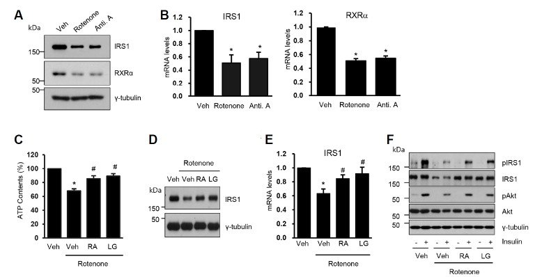
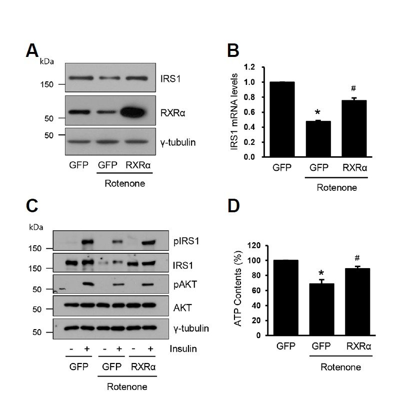
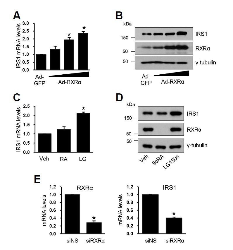
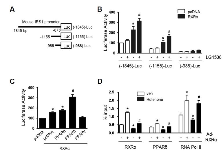
Fig. 1.RXRα agonists restore mitochondrial function and IRS transcription impaired by rotenone. C2C12 myoblasts were differentiated to myotubes and treated with rotenone (3 μM) or antimycin A (10 μM) for 24 h. (A) Cells were lysed and then Western blot analyses were performed with the specific antibodies. (B) Total RNAs were isolated and real time quantitative PCR was performed. The mRNA levels of each gene were normalized by that of GAPDH. The value of Veh was set to 1 and the others were expressed as the relatives to that. The data are the means ± SEM of 3 experiments. *, P < 0.05 vs. Veh. (C-F) C2C12 myotubes were treated with rotenone (3 μM) for 24 h and then the media was replaced to the fresh media containing DMSO (Veh), 9cRA (5 μM) or LG1506 (2 μM) for 18 h. (C) Cells were lysed and ATP contents were measured (n = 6). *, P < 0.05 vs. Veh; #, P < 0.05 vs. rotenone only. (D) Cell lysates were subjected to Western blot analysis. (E) The IRS1 mRNA levels were measured by real time PCR (n = 5). *, P < 0.05 vs. Veh; #, P < 0.05 vs. rotenone only. (F) Insulin (100 nM) was added 15 min before harvesting. Cell lysates were subjected to the Western blot analysis.Fig. 2.RXRα overexpression prevents rotenone-induced suppression of IRS1 transcription. C2C12 myotubes were infected with Ad-RXRα (50 MOI) for 48 h and then treated with rotenone for an additional 24 h. (A) Western blot analysis and (B) real-time PCR were performed (n = 6). P < 0.05 vs. Ad-GFP; #, P < 0.05 vs. Ad-GFP with rotenone. (C) Insulin was treated for 15 min before harvesting and western blot analysis was performed. (D) Cellular ATP contents were measured (n = 7). *, P < 0.05 vs. Ad-GFP; #, P < 0.05 vs. Ad-GFP with rotenone.Fig. 3.RXRα directly regulates IRS1 transcription. (A, B) C2C12 myotubes were infected with Ad-RXRα in different dosages (10, 30 and 50 MOI) for 48 h. Cell lysates were subjected to real time PCR (A) and western blot analysis (B). The mRNA levels of control cells (Ad-GFP infected) was set to 1 and the other values were expressed as the relatives to that (n = 4). *, P < 0.05 vs. Ad-GFP infected cells. (C, D) C2C12 myotubes were treated with 9cRA (5 μM) or LG1506 (2 μM) for 24 h, and then real time PCR (C) and western blot analysis (D) were performed (n = 5). *, P < 0.05 vs. Veh. (E) C2C12 myotubes were transfected with siNS or siRXR (50 nM). Total RNAs were prepared 3 days after siRNA transfection and then real time PCR was performed (n = 3). *, P < 0.05 vs. siNS.Fig. 4.RXRα directly binds to the IRS1 promoter. (A) A diagram of three different lengths of IRS1 promoter-luciferase constructs, IRS1 (-1845)-Luc, IRS1 (-1155)-Luc and IRS1 (-998)-Luc. (B) COS7 cells were transfected with one of the IRS1 promoter-Luc constructs and RXRα expression vector (pcDNARXRα). Three hours after the transfection, cells were treated with LG1506 (2 μM) for an additional 18 h. Luciferase activity of the cells transfected with IRS1 (-1845)-Luc was set to 100 and the other values were expressed as the relatives to that (n = 5). *, P < 0.05 vs pcDNA without LG1506 for each construct. #, P < 0.05 vs. pcDNA-RXRα without LG1506. (C) COS7 cells were transfected with IRS1 (-1155) Luc, expression vectors for RXRα, PPARα, PPARδ or PPARγ. Luci ferase activity of the cells transfected with IRS1 (-1155)-Luc without RXRα or PPAR expression vectors was set to 100 and the others were expressed as the relatives to that (n = 4). *, P < 0.05 vs. cells transfected with IRS1 (-1155)-Luc only. #, P < 0.05 vs. cells transfected with IRS1 (-1155)-Luc and pcDNA-RXRα (D) C2C12 myotubes were infected with Ad-RXRα (50 MOI) for 48 h, and then treated with rotenone for an additional 24 h. Cells were subjected to ChIP. Data are the means ± SEM (n = 3). *, P < 0.05 vs. Ad-GFP infected cells not treated with rotenone. #, P < 0.05 vs. Ad-GFP infected cells treated with rotenone.