Department of Internal Medicine, Seoul National University Boramae Medical Center, Seoul, 07061, South Korea.
Department of Laboratory Medicine, College of Medicine, The Catholic University of Korea, Seoul, 06591, South Korea.
Catholic Genetic Laboratory Center, Seoul St. Mary's Hospital, College of Medicine, The Catholic University of Korea, Seoul, 06591, South Korea.
Department of Neurosurgery, College of Medicine, Korea University, Seoul, 02841, South Korea.
Department of Integrated Biomedical and Life Sciences, College of Health Science, Korea University, Seoul, 02841, South Korea.
Center for Genomic Research and Development, Korea University Anam Hospital, Seoul, 02841, South Korea.
Department of Thoracic & Cardiovascular surgery, College of Medicine, Korea University, Seoul, 02841, South Korea.
Department of Medical Science, Soonchunhyang University, Asan-si, Chungcheongnam-do, 31538, South Korea.
Department of Pre-Medicine, College of Medicine, Hanyang University, Seoul, 04763, South Korea.
Hanyang Institute of Bioscience and Biotechnology, Hanyang University, Seoul, 04763, South Korea.
Division of Hepato-Biliary and Pancreatic Surgery, Department of Surgery, BK21 Project, Asan Medical Center, University of Ulsan College of Medicine, Seoul, 05505, South Korea.
Department of Genetics, College of Medicine, Hanyang University, Seoul, 04763, South Korea.
Graduate School of Biomedical Science and Engineering, Hanyang University, Seoul, 04763, South Korea.
Abstract
In this study, we present the development of the Mutation tagging by CRISPR-based Ultra-precise Targeted Elimination in Sequencing (MUTE-Seq) method. We engineered a highly precise advanced-fidelity FnCas9 variant, named FnCas9-AF2, to effectively discriminate single-base mismatches at all positions of the single guide RNA (sgRNA) target sequences. FnCas9-AF2 exhibited significantly lower off-target effects compared to existing high-fidelity CRISPR-Cas9 variants. MUTE-Seq leverages FnCas9-AF2 for the enrichment of mutant DNA through the exclusive cleavage of perfectly matched wild-type DNA, allowing for sensitive detection of low-frequency cancer-associated mutant alleles. MUTE-Seq enabled sensitive monitoring of minimal residual disease (MRD) from the bone marrow of patients with Acute Myeloid Leukemia (AML). Furthermore, MUTE-Seq was applied in a multiplexed manner on cell-free DNA (cfDNA) from patients diagnosed with non-small cell lung cancer (NSCLC) and pancreatic cancer. This approach demonstrated a significant improvement in the sensitivity of simultaneous mutant detection and highlighted its clinical utility for early-stage cancer patients with extremely low levels of circulating tumor DNA (ctDNA). We anticipate that the FnCas9-AF2-based MUTE-Seq could offer a valuable clinical tool to facilitate improved molecular diagnosis, prognosis evaluation, and treatment planning for cancers in various stages.
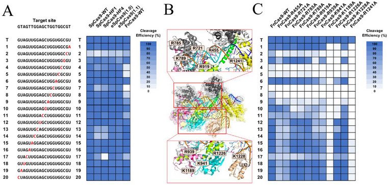
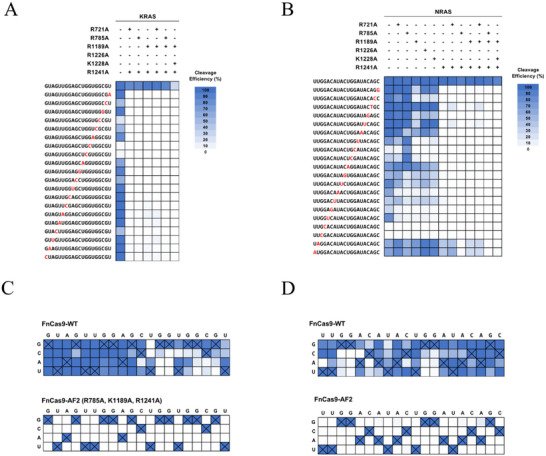
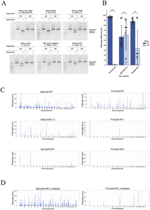
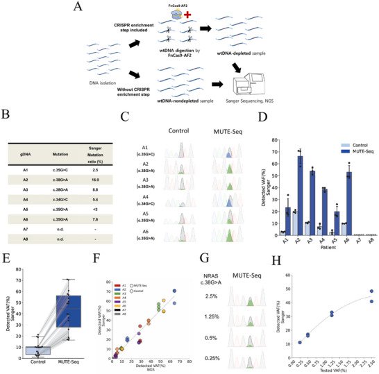
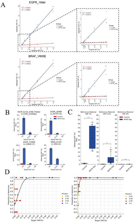
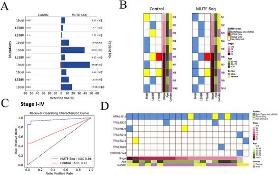
Figure 1Enhancing the Base Mismatch Sensitivity and Specificity of FnCas9 Through Protein Engineering. A) Sequences of sgRNAs targeting a KRAS site with a single mismatch and in vitro cleavage efficiencies of SpCas9‐WT, SpCas9‐HF1, SpCas9‐HF4, eSpCas9(1.0), eSpCas9(1.1), and FnCas9‐WT proteins. B) Positions of amino acids within the crystal structure of FnCas9 (PDB 5B2O) that were changed in the 11 highly precise mutants; six with modified residues in the Rec and PLL domain (R455, R785, K721, K789, R919, and R1241) that interacted with the phosphate backbone of the target DNA strand, and five with modified residues in the HNH, RuvC and PLL domain (R939, K941, K1189, R1226, K1228) that interacted with the phosphate backbone of the non‐target DNA strand. C) Quantification of target cleavage efficiencies of the 11 FnCas9 variants with single amino acid substitution. (T: On‐target sequence; Numbers: Single‐base mismatch positions counted from PAM).Figure 2Comparative Cleavage Efficiencies and Specificities of FnCas9 Variants against Single‐Mismatched sgRNA Targets. A) Assessment of specificity of FnCas9 variants with single, double, and triple mutations. Heatmaps show cleavage efficiencies of FnCas9 variants with single‐mismatched sgRNAs against a KRAS target. B. Specificity assessment of FnCas9 variants against an NRAS target. C. Cleavage efficiencies of FnCas9 and FnCas9‐AF2 (R785A, K1189A, R1241A triple mutant) for sgRNAs targeting the KRAS with single‐base mismatches at all 20 nucleotide positions. D. Cleavage rates of FnCas9 and FnCas9‐AF2 for sgRNAs with single‐base mismatches at all 20 positions of the NRAS target. The perfect base‐pair between the sgRNA and the target DNA in each nucleotide position is marked by the cross mark. The cleavage rates are shown in shades of blue.Figure 3Genome‐wide assessment of effective discrimination of mutations by FnCas9 and SpCas9 Variants. A) In vitro cleavage efficiency of FnCas9‐AF2 against wild‐type and mutant DNA targets. SNVs (EGFR c.2573 T>G, EGFR c.2369 C>T, EGFR c.2389 T>A, KRAS c.35 G>A, MET c.3028+1 G>A) and an indel variant (EGFR c.2236_2250 del) were assessed. Cleavage of wild‐type DNA by FnCas9‐AF2 resulted in two distinct DNA fragments, while mutant‐type DNA remained intact, illustrating FnCas9‐AF2's specificity for wild‐type over mutant DNA targets. B) Comparative in vitro cleavage efficiencies of FnCas9‐AF2, eSpCas9(1.1), and SpCas9‐HF4. While SpCas9 variants show variable levels of cleavage efficiencies, FnCas9‐AF2 demonstrates consistent, high cleavage rates of wild‐type DNA (96.84% in average) and markedly low cleavage rates for mutant DNA (0.21% in average). Data for independent biological replicates (n = 3) are depicted by black dots, and error bars represent SEM. C) Genome‐wide analyses of off‐target cleavage sites were conducted with Digenome‐seq for SpCas9‐WT, eSpCas9(1.1), SpCas9‐HF4, FnCas9‐WT, FnCas9‐AF1, and FnCas9‐AF2, using NRAS‐targeting sgRNA. The graphs show the genomic locations of off‐target sites cleaved by the Cas9 variants. D) Genome‐wide multiplex analyses of off‐target cleavage sites were conducted with Multiplex Digenome‐seq for SpCas9‐HF4 and FnCas9‐AF2, using gRNAs targeting five genomic locations (FANCF, NRAS, AAVS1, HBB, DMD). The graphs show the genomic locations of off‐target sites cleaved by the Cas9 variants.Figure 4Evaluation of MUTE‐Seq and Clinical Validation in AML patients. A) Schematic diagram demonstrating the process of MUTE‐Seq. This includes the in vitro digestion of wtDNA using FnCas9‐AF2, and the subsequent analysis of the enriched mutant DNA via Sanger and Next Generation Sequencing (NGS). B) Table of eight clinical samples showing NRAS mutations and corresponding Sanger sequencing VAF(%) calculated as the “Mutant peak height/(Mutant peak height + Wild‐type height) x 100(%)”. C) Sanger sequencing chromatograms of eight clinical samples, both before (control) and after MUTE‐Seq treatment, demonstrating the presence of NRAS mutations. D) VAFs from Sanger sequencing of eight clinical samples, before and after MUTE‐Seq treatment. The error bars represent the SEM from three independent experiments. E) Box plot showing the VAFs NRAS mutation of all patients before and after MUTE‐Seq treatment, respectively. Gray lines demonstrate pairing the same patient samples before and after MUTE‐Seq treatment. F) Scatter plot showing the correlation between Sanger sequencing VAFs and NGS VAFs. Circles and diamonds represent the MUTE‐Seq and control group, respectively. The line represents the linear regression (R2 = 0.958). G) Sanger sequencing chromatograms after enrichment for reference material created by mixing patient sample with the NRAS c.38G>A mutation and wild‐type gDNA to reach initial mutant ratios of 2.5, 1.25, 0.5, and 0.25%. H) Scatter plot for original VAF and detected VAF (R2 = 0.977).Figure 5Enhancing the Detection Sensitivity of Low‐frequency Mutations using Multiplexed MUTE‐Seq. A) Detected VAFs from diluted plasmid EGFR exon 19 deletion (top) and BRAF V600E (bottom) DNA samples of 0.5 to 0.005% VAFs for three MUTE‐Seq groups and three control groups. The linear regression line of best fit, along with the regression coefficient, is presented. Right side panels are magnified graphs focusing on the original VAFs ranging from 0.1 to 0.005% with a separate linear regression line of best fit and a regression coefficient. The dotted line indicates 0.1% VAF. B) Detected VAFs using NGS for control and multiplexed MUTE‐Seq on cfDNA reference material containing EGFR exon 19del, EGFR T790M, EGFR L858R, and KRAS G12D mutations of 1, 0.1, and 0% (n = 3, respectively). C) Box‐and‐whisker plot displaying the comparison of combined VAFs detected by control and MUTE‐seq. D) Probability‐of‐detection curves for 5 ng (left) and 50 ng (right) DNA inputs with probit regression fits (solid lines). The estimated 95% limits of detection were 0.15% VAF [95% CI, 0.08–0.22%] for 5 ng and 0.034% VAF [95% CI, 0.018–0.051%] for 50 ng.Figure 6Evaluation of Multiplexed MUTE‐Seq and Clinical Validation in Non‐Small Cell Lung Cancer (NSCLC) and Pancreatic Cancer Patients. A) VAF (%) of EGFR variants in cfDNA of NSCLC patients in control and MUTE‐Seq groups. B) Detection of EGFR mutations in control and MUTE‐Seq (EGFR Exon 19 deletion, EGFR T790M, and EGFR L858R) in tissue and blood specimens from 10 NSCLC patients. C) ROC analysis between tissue and blood samples from 10 NSCLC patients in relation to EGFR mutations. Lines colored in blue and red denote the ROC curves of multiplexed MUTE‐seq and control, respectively. D) Detection of pancreatic cancer‐associated mutations in tissue and cfDNA of 20 pancreatic cancer patients with MUTE‐Seq.
그림 출처: PubMed Central 공개 논문 (PMC12651102)
Conflict of interest statement
S.Y. filed patent applications based on this study.
그 밖의 정보
출판 형태
Journal Article, Research Support, Non-U.S. Gov't
저널
Advanced materials (Deerfield Beach, Fla.) · eISSN 1521-4095
Cell-Free Nucleic Acids · RNA, Guide, CRISPR-Cas Systems
연구비
National Institute for International Education 2021R1A6A1A03040260 · National Institute for International Education RS-2023-00260529 · Korea National Institute of Health RS-2024-00435385 · Korea Medical Device Development Fund RS-2021-KD000007 · National Research Foundation of Korea RS-2021-NR056589 · National Research Foundation of Korea RS-2023-00260529 · National Research Foundation of Korea RS-2023-00262309 · National Research Foundation of Korea RS-2025-02218918 · Korea Ministry of Health & Welfare RS-2022-KH129266 · Korea University K2125811