
Eur Radiol. 2026;36(5):4177-4186.
Enhancing vertebral fracture prediction using multitask deep learning computed tomography imaging of bone and muscle.
제1저자 공성혜, 최새미 · 교신저자 박성배, 김정희
소속 (10)
- Department of Internal Medicine, Seoul National University Bundang Hospital, Bundang, Gyeonggi-do, Republic of Korea.
- Department of Internal Medicine, Seoul National University College of Medicine, Seoul, Republic of Korea.
- Kim Jaechul Graduate School of AI, Korea Advanced Institute of Science and Technology, Daejeon, Republic of Korea.
- Letsur Inc., Seoul, Republic of Korea.
- Department of Medical Device Development, Seoul National University College of Medicine, Seoul, Republic of Korea. ddolbae01@snu.ac.kr.
- Department of Neurosurgery, Seoul National University Boramae Hospital, Seoul, Republic of Korea. ddolbae01@snu.ac.kr.
- Department of Internal Medicine, Seoul National University Hospital, Seoul, Republic of Korea.
- Department of Internal Medicine, Seoul National University College of Medicine, Seoul, Republic of Korea. jhee1@snu.ac.kr.
- Department of Internal Medicine, Seoul National University Hospital, Seoul, Republic of Korea. jhee1@snu.ac.kr.
- Department of Internal Medicine, Seoul National University Boramae Hospital, Seoul, Republic of Korea.
Abstract
OBJECTIVES To develop and externally validate a computed tomography (CT)-based multitask learning model to predict fracture risk.
MATERIALS AND METHODS This study was conducted in two parts, using a multitasking learning approach. We developed a cross-sectional vertebral fracture (VF) detection model using abdominal CT scans of 2553 patients aged 50-80 years. Then, we leveraged this detection model within a multitask learning framework to develop a longitudinal VF prediction model over a 5-year follow-up period. External testing was performed on 1506 patients from two independent hospitals. The performance was compared between the single-task and multitask models, bone-only and bone+muscle images, and image-only and clinical models.
RESULTS For the cross-sectional fracture detection model, the mean age of the patients was 76.2 years, and 66.7% were female. In the classification task for detection of VF, the model using both bone and muscle showed an area under the receiver operating characteristic curve (AUROC) of 0.82 in the development set and 0.80 in the external test sets. Using multitask learning, the bone + muscle image model showed a c-index of 0.68 and had superior performance than the bone-only model in the external test set for 2-year, 3-year, and 5-year AUROCs (0.79 vs. 0.75, 0.71 vs. 0.68, and 0.71 vs. 0.68, respectively, all p < 0.01). Also, the multitask model significantly outperformed the Fracture Risk Assessment Tool (FRAX) (c-index: 0.68 vs. 0.66, p < 0.01).
CONCLUSION The CT-based multitask learning model integrating both bone and muscle data showed superior predictive performance for VFs compared with models using bone images only and traditional clinical models.
KEY POINTS Question Vertebral fracture risk remains underestimated in many individuals undergoing CT scans for other reasons, highlighting the need for improved opportunistic prediction tools. Findings A multitask deep learning model integrating both bone and muscle features from CT scans demonstrated superior performance compared to bone-only and traditional clinical models, including FRAX. Clinical relevance The proposed model enables accurate vertebral fracture risk prediction using routinely acquired CT scans, facilitating early identification and intervention without the need for additional tests.
© 2025. The Author(s).
Keywords
Deep learningFractureFracture risk assessmentLongitudinal cohortPrediction
Figures
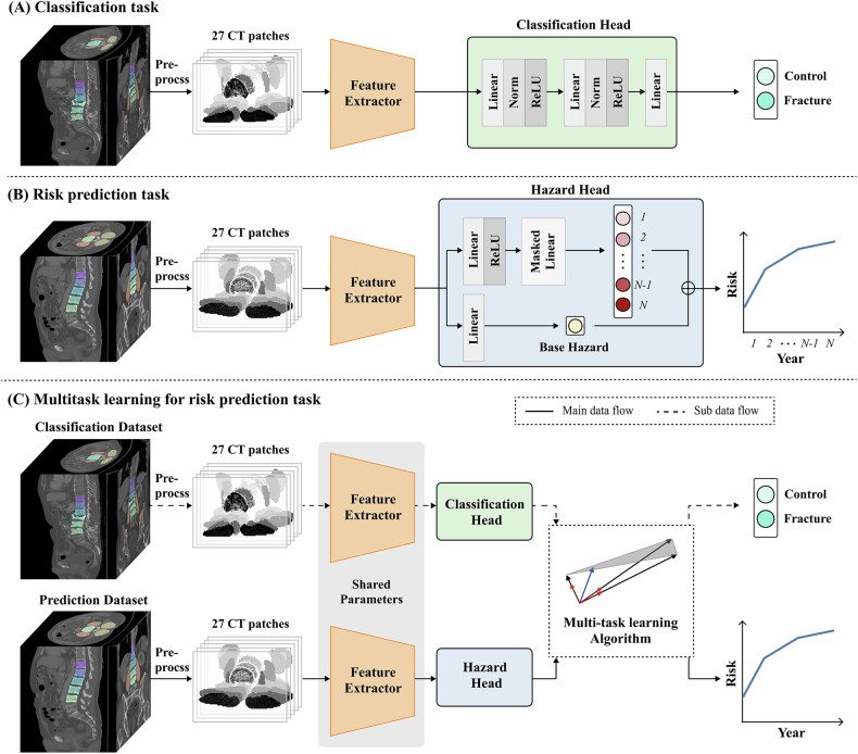
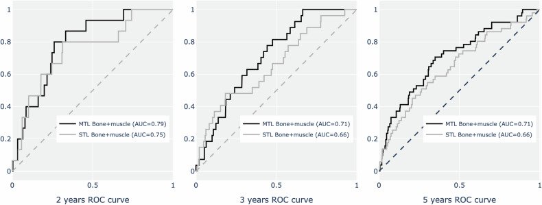
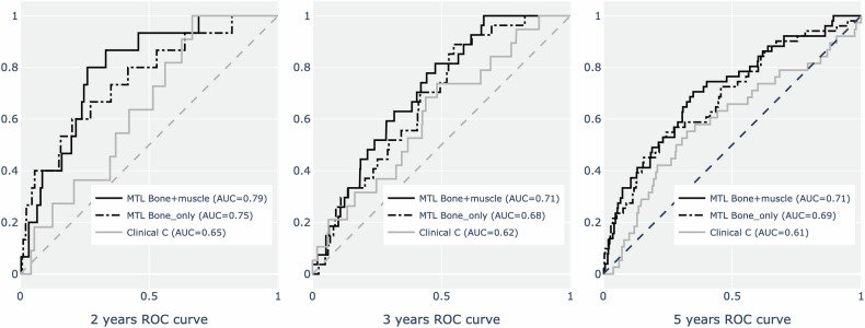
그림 출처: PubMed Central 공개 논문 (PMC13086793)
Conflict of interest statement
Compliance with ethical standards. Guarantor: The scientific guarantor of this publication is Jung Hee Kim. Conflict of interest: The authors of this manuscript declare no relationships with any companies, whose products or services may be related to the subject matter of the article. Statistics and biometry: No complex statistical methods were necessary for this paper. Informed consent: Written informed consent was waived by the Institutional Review Board. Ethical approval: Institutional Review Board approval was obtained. Study subjects or cohorts overlap: Some study subjects or cohorts have not been previously reported. Methodology: Retrospective Observational Multicenter study
그 밖의 정보
- 출판 형태
- Journal Article
- 저널
- European radiology · eISSN 1432-1084
- 전자 출판
- 2025-12-01
- MeSH
- Humans · Female · Aged · Tomography, X-Ray Computed* / methods · Deep Learning* · Male · Middle Aged · Spinal Fractures* / diagnostic imaging · Aged, 80 and over · Predictive Value of Tests · Cross-Sectional Studies · Bone and Bones / diagnostic imaging
- 연구비
- National Research Foundation of Korea 2020R1A2C2011587, 2021R1A2C2003410
- 참고문헌
- 30편