
Endocrinol Metab (Seoul). 2021;36(2):447-454.
DEHP Down-Regulates Tshr Gene Expression in Rat Thyroid Tissues and FRTL-5 Rat Thyrocytes: A Potential Mechanism of Thyroid Disruption.
제1저자 김민주 · 교신저자 박영주, 오병철
소속 (7)
- Seoul National University Hospital Healthcare System Gangnam Center, Seoul, Korea.
- Department of Internal Medicine, Seoul National University College of Medicine, Seoul, Korea.
- Department of Internal Medicine, CHA Bundang Medical Center, CHA University, Seongnam, Korea.
- Department of Physiology, Lee Gil Ya Cancer and Diabetes Institute, Gachon University College of Medicine, Incheon, Korea.
- Graduate School of Public Health, Seoul National University, Seoul, Korea.
- Department of Environmental Science and Institute of Biomedical Studies, Baylor University, Waco, TX, USA.
- Department of Molecular Medicine and Biopharmaceutical Sciences, Graduate School of Convergence Science and Technology, Seoul National University, Seoul, Korea.
Abstract
BACKGROUND Di-2-ethylhexyl phthalate (DEHP) is known to disrupt thyroid hormonal status. However, the underlying molecular mechanism of this disruption is unclear. Therefore, we investigated the direct effects of DEHP on the thyroid gland.
METHODS DEHP (vehicle, 50 mg/kg, and 500 mg/kg) was administered to Sprague-Dawley rats for 2 weeks. The expression of the thyroid hormone synthesis pathway in rat thyroid tissues was analyzed through RNA sequencing analysis, quantitative reverse transcription-polymerase chain reaction (RT-PCR), and immunohistochemical (IHC) staining. DEHP was treated to FRTL-5 rat thyroid cells, and an RT-PCR analysis was performed. A reporter gene assay containing the promoter of thyroid stimulating hormone receptor (TSHR) in Nthy-ori 3-1 human thyroid cells was constructed, and luciferase activity was determined.
RESULTS After DEHP treatment, the free thyroxine (T4) and total T4 levels in rats significantly decreased. RNA sequencing analysis of rat thyroid tissues showed little difference between vehicle and DEHP groups. In the RT-PCR analysis, Tshr expression was significantly lower in both DEHP groups (50 and 500 mg/kg) compared to that in the vehicle group, and IHC staining showed that TSHR expression in the 50 mg/kg DEHP group significantly decreased. DEHP treatment to FRTL-5 cells significantly down-regulated Tshr expression. DEHP treatment also reduced luciferase activity in a reporter gene assay for TSHR.
CONCLUSION Although overall genetic changes in the thyroid hormone synthesis pathway are not clear, DEHP exposure could significantly down-regulate Tshr expression in thyroid glands. Down-regulation of Tshr gene appears to be one of potential mechanisms of thyroid disruption by DEHP exposure.
Keywords
Diethylhexyl phthalateEndocrine disruptorsReceptors, thyrotropinThyroid gland
Figures
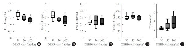
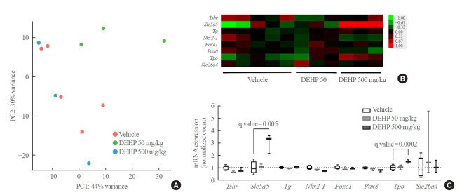
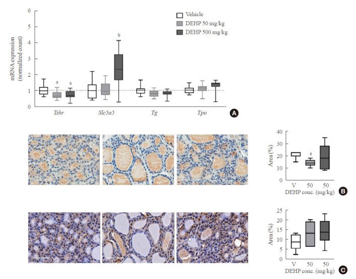
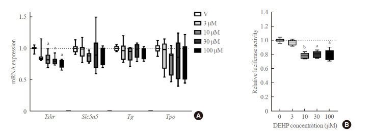
그림 출처: PubMed Central 공개 논문 (PMC8090463)
Conflict of interest statement
CONFLICTS OF INTEREST. No potential conflict of interest relevant to this article was reported.
그 밖의 정보
- 출판 형태
- Journal Article, Research Support, Non-U.S. Gov't
- 저널
- Endocrinology and metabolism (Seoul, Korea) · eISSN 2093-5978
- 전자 출판
- 2021-03-31
- MeSH
- Animals · Diethylhexyl Phthalate* / metabolism / toxicity · Gene Expression · Rats · Rats, Sprague-Dawley · Receptors, Thyrotropin / genetics · Thyroid Epithelial Cells* / metabolism · Thyroid Gland / metabolism
- Substances
- Receptors, Thyrotropin · Diethylhexyl Phthalate
- 참고문헌
- 25편