
J Korean Med Sci. 2015;30(6):716-24.
Three dimensional quantitative coronary angiography can detect reliably ischemic coronary lesions based on fractional flow reserve.
제1저자 정우영 · 교신저자 Lerman A
소속 (5)
- Devision of Cardiology, Department of Internal Medicine, Boramae Medical Center, Seoul National University, College of Medicine, Seoul, Korea.
- Department of Cardiology, Ajou University School of Medicine, Suwon, Korea.
- Division of Cardiology, Department of Internal Medicine, School of Medicine, Dankook University, Cheonan, Korea.
- Department of Cardiovascular Medicine, Wakayama Medical University, Wakayama, Japan.
- Division of Cardiovascular Disease, Mayo Clinic, Rochester, MN, USA.
Abstract
Conventional coronary angiography (CAG) has limitations in evaluating lesions producing ischemia. Three dimensional quantitative coronary angiography (3D-QCA) shows reconstructed images of CAG using computer based algorithm, the Cardio-op B system (Paieon Medical, Rosh Ha'ayin, Israel). The aim of this study was to evaluate whether 3D-QCA can reliably predict ischemia assessed by myocardial fractional flow reserve (FFR) < 0.80. 3D-QCA images were reconstructed from CAG which also were evaluated with FFR to assess ischemia. Minimal luminal diameter (MLD), percent diameter stenosis (%DS), minimal luminal area (MLA), and percent area stenosis (%AS) were obtained. The results of 3D-QCA and FFR were compared. A total of 266 patients was enrolled for the present study. FFR for all lesions ranged from 0.57 to 1.00 (0.85 ± 0.09). Measurement of MLD, %DS, MLA, and %AS all were significantly correlated with FFR (r = 0.569, 0609, 0.569, 0.670, respectively, all P < 0.001). In lesions with MLA < 4.0 mm(2), %AS of more than 65.5% had a 80% sensitivity and a 83% specificity to predict FFR < 0.80 (area under curve, AUC was 0.878). 3D-QCA can reliably predict coronary lesions producing ischemia and may be used to guide therapeutic approach for coronary artery disease.
Keywords
Coronary AngiographyCoronary Artery DiseaseMyocardial Ischemia
Figures


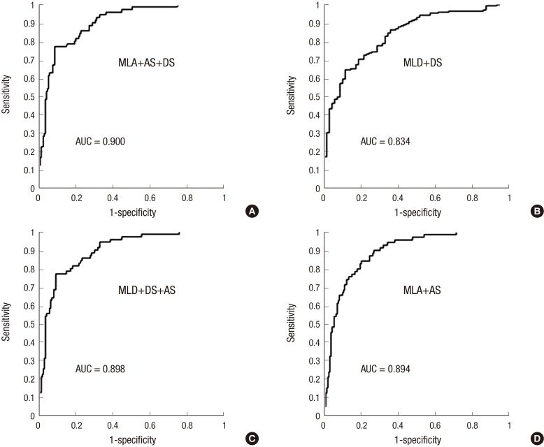
그림 출처: PubMed Central 공개 논문 (PMC4444471)
Conflict of interest statement
DISCLOSURE: The authors have no conflicts of interest to disclose.
그 밖의 정보
- 출판 형태
- Journal Article
- 저널
- Journal of Korean medical science · eISSN 1598-6357
- 전자 출판
- 2015-05-13
- MeSH
- Aged · Coronary Angiography / methods · Coronary Circulation · Coronary Stenosis / diagnostic imaging / etiology / physiopathology · Female · Fractional Flow Reserve, Myocardial* · Humans · Imaging, Three-Dimensional / methods · Male · Myocardial Ischemia / complications / diagnostic imaging / physiopathology · Radiographic Image Enhancement / methods · Radiographic Image Interpretation, Computer-Assisted / methods · Reproducibility of Results · Sensitivity and Specificity
- 참고문헌
- 26편