Strategic Center of Cell & Bio Therapy, Seoul National University Hospital, Seoul 03080, Republic of Korea; Department of Molecular Medicine and Biopharmaceutical Sciences, Graduate School of Convergence Science and Technology and College of Medicine or College of Pharmacy, Seoul National University, Seoul 03080, Republic of Korea.
Strategic Center of Cell & Bio Therapy, Seoul National University Hospital, Seoul 03080, Republic of Korea.
Division of Cardiology, Department of Internal Medicine, Seoul National University Hospital, Seoul 03080, Republic of Korea. Electronic address: hyunjaicho@snu.ac.kr.
Strategic Center of Cell & Bio Therapy, Seoul National University Hospital, Seoul 03080, Republic of Korea; Department of Molecular Medicine and Biopharmaceutical Sciences, Graduate School of Convergence Science and Technology and College of Medicine or College of Pharmacy, Seoul National University, Seoul 03080, Republic of Korea. Electronic address: hyosoo@snu.ac.kr.
Abstract
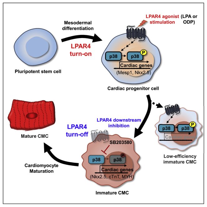
Efficient differentiation of pluripotent stem cells (PSCs) into cardiac cells is essential for the development of new therapeutic modalities to repair damaged heart tissue. We identified a novel cell surface marker, the G protein-coupled receptor lysophosphatidic acid receptor 4 (LPAR4), specific to cardiac progenitor cells (CPCs) and determined its functional significance and therapeutic potential. During in vitro differentiation of mouse and human PSCs toward cardiac lineage, LPAR4 expression peaked after 3-7 days of differentiation in cardiac progenitors and then declined. In vivo, LPAR4 was specifically expressed in the early stage of embryonal heart development, and as development progressed, LPAR4 expression decreased and was non-specifically distributed. We identified the effective agonist octadecenyl phosphate and a p38 MAPK blocker as the downstream signal blocker. Sequential stimulation and inhibition of LPAR4 using these agents enhanced the in vitro efficiency of cardiac differentiation from mouse and human PSCs. Importantly, in vivo, this sequential stimulation and inhibition of LPAR4 reduced the infarct size and rescued heart dysfunction in mice. In conclusion, LPAR4 is a novel CPC marker transiently expressed only in heart during embryo development. Modulation of LPAR4-positive cells may be a promising strategy for repairing myocardium after myocardial infarction.
G protein-coupled receptorGPCRcell therapydifferentiationmyocardial infarctionstem cells
Figures
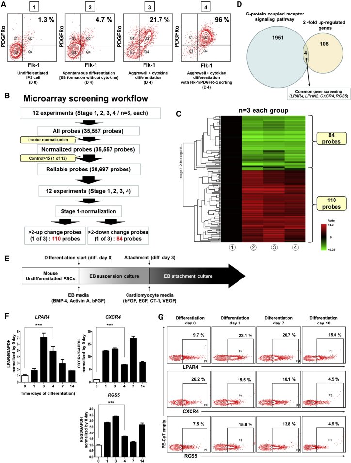
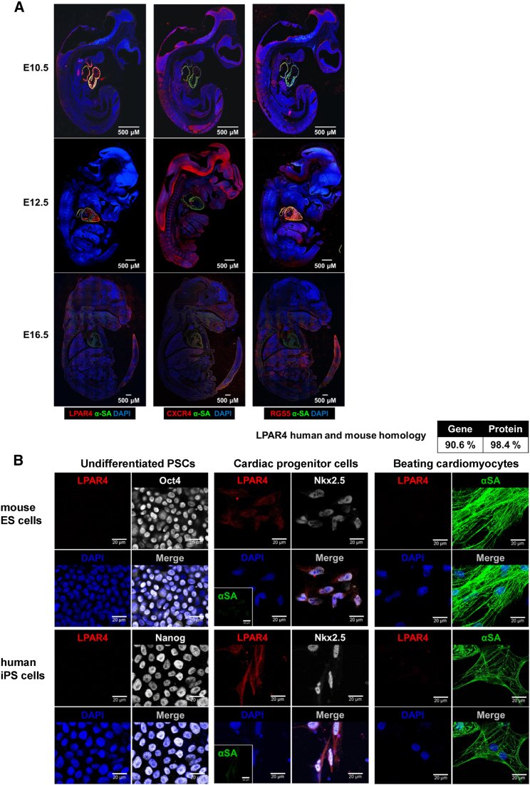
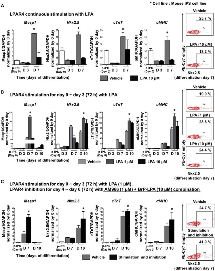
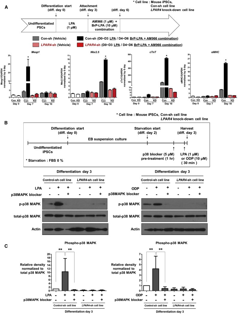
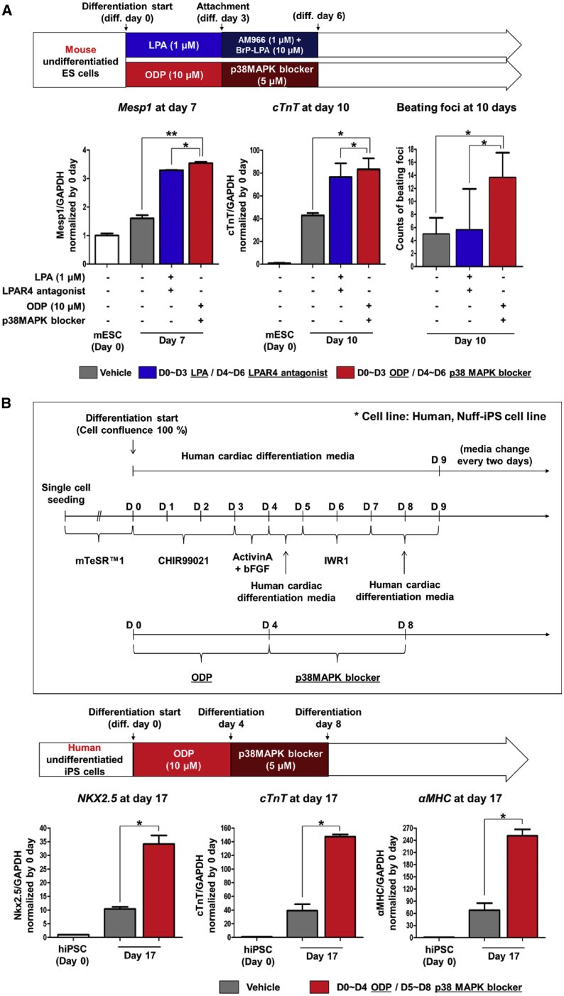
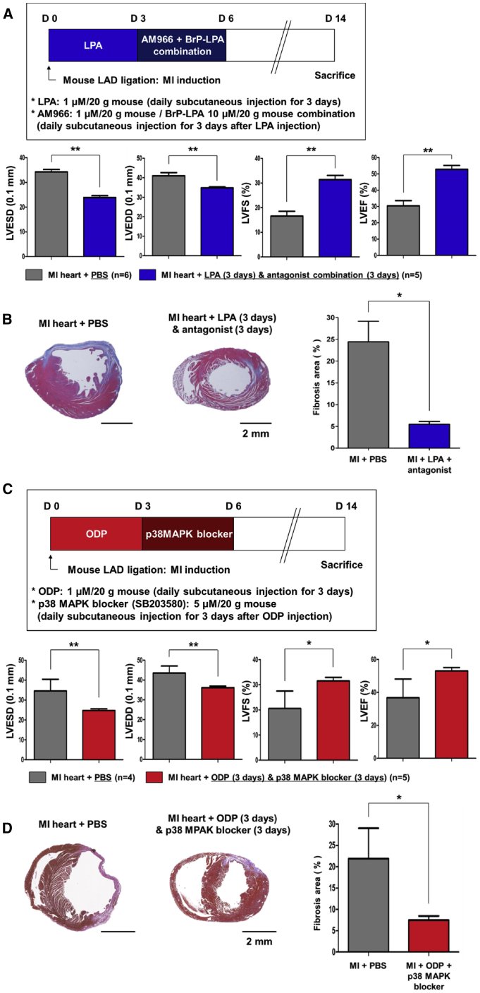
Figure 1Identification and Expression of a New Cardiac Progenitor-Specific Marker (A) FACS analysis with the well-known cardiac progenitor markers Flk-1 and PDGFRα in undifferentiated induced pluripotent stem cells (iPSCs, group 1) and three cell lines at different stages of day 4-differentiation: group 2, spontaneously differentiated cells; group 3, cells differentiated by the established cardiac differentiation protocol; group 4, cells differentiated by the established cardiac differentiation protocol and sorted by Flk-1 and PDGFRα. (B) Microarray screening workflow for the four cell groups (n = 3). (C) Heatmap data from the microarray screening workflow. (D) Four candidate markers showing 2-fold upregulated expression are all G-protein–coupled receptors (GPCR): lysophosphatidic acid receptor 4 (LPAR4), Latrophilin 2 (LPHN2), chemokine (C-X-C motif) receptor 4 (CXCR4), and a regulator of G-protein signaling 5 (RGS5). (E) Schematic representation of the established cardiac differentiation protocol. (F) The real-time PCR analysis of the mRNA expression levels of the three candidates during cardiac differentiation. Error bars represent SEM, ∗∗∗p < 0.001, unpaired t test. n = 3 biological replicates. (G) FACS analysis of the protein expression levels of the three candidates during cardiac differentiation. All experiments were conducted at least in triplicate.Figure 2Transient Expression Pattern of LPAR4 (A) Immunofluorescence analysis during development at embryonic day 10.5, 12.5, and 16.5 (E10.5, E12.5, and E16.5). Red, CXCR4, RGS5, and LPAR4; green, αSA; DAPI, nuclei. Scale bar, 500 μm. (B) Immunofluorescence (IF) analysis of the correlation between representative cardiac progenitor markers and LPAR4 during differentiation from mouse ESCs and human iPSCs to CMCs. Red, LPAR4; green, αSA; white, Oct4, Nanog, and Nkx2.5; DAPI, nuclei. Scale bar, 20 μm. All experiments were conducted at least in triplicate.Figure 3Protocol to Improve Cardiac Differentiation by LPAR4 Regulation (A) Real-time PCR and FACS analyses confirmed the expression of well-known cardiac progenitor markers when LPAR4 was continuously stimulated by treatment with 10 μM LPA by the established cardiac differentiation protocol. Real-time PCR analysis at cardiac differentiation day 3 and day 7, FACS analysis at day 7. (B) LPA stimulation during the early differentiation stage of the established cardiac differentiation protocol. LPAR4 was stimulated by 1 μM and 10 μM LPA for 3 days, and the results were compared to those of the untreated group. (C) LPAR4 was stimulated by 1 μM LPA for the first 3 days of differentiation and then inhibited by a combination of the antagonists AM966 and BrP-LPA for the next 3 days, and the results were compared with those of the untreated group. Statistical analyses were performed using one-way ANOVA (Newman-Keuls). ∗p < 0.01. All experiments were conducted at least in triplicate.Figure 4Identification of LPAR4 Downstream Signaling Molecules (A) Expression of cardiac progenitor markers in the LPAR4-knockdown cell line and control cell line by real-time PCR at cardiac differentiation day 7 and day 10; the LPAR4-knockdown cell line did not differentiate into CMCs. Statistical analyses were performed using one-way ANOVA (Newman–Keuls). ∗p < 0.01. (B) The effects of LPA and ODP were confirmed by western blotting in the control cell line and LPAR4-knockdown cell line at cardiac differentiation day 3. Both cell lines were starved for 1 day and treated with LPA for 30 min. The cells were treated with p38 MAPK 30 min before LPA treatment. All experiments were conducted at least in triplicate. (C) Quantification of western blot from Figure 4B. Error bars represent the mean of four independent experiments. ∗p < 0.01.Figure 5Cardiac Differentiation Protocol Using Sequential Stimulation and Then Inhibition of LPAR4 in Mouse ESCs and Human iPSCs (A) To maximize the cardiac differentiation efficiency, LPA or ODP was used as LPAR4 stimulants. LPAR4 antagonist combination (BrP-LPA and AM966) or p38 MAPK blocker was used as LPAR4 inhibitors. Mouse ESCs were differentiated into CMCs and confirmed using the cardiac lineage marker Mesp1 at cardiac differentiation day 7 and cTnT at cardiac differentiation day 10 by real-time PCR. Besides, beating CMCs were counted (displayed as a bar graph) to confirm the cardiac differentiation efficiency. (B) The human cardiac differentiation protocol is shown in a schematic figure (upper panel). The effects of ODP and the p38 MAPK blocker on the differentiation efficiency of human iPSCs into CMCs were evaluated. The cardiac lineage markers were analyzed by real-time PCR at cardiac differentiation day 17 (lower panel). Statistical analyses were performed using one-way ANOVA (Newman-Keuls). ∗∗∗p < 0.001. All experiments were conducted at least in triplicate.Figure 6The Therapeutic Effect of Sequential Stimulation and then Inhibition of LPAR4 Signaling in a Mouse MI Model (A) Schematic representation of subcutaneously injected with the LPAR4 non-specific agonist LPA (1 μM/20 g) and antagonist combination (AM966 [1 μM/20 g] + BrP-LPA [10 μM/20 g]) in the mouse MI model. Echocardiography (MI + PBS group, n = 6; MI + LPA [3 days] + antagonist [3 days] group, n = 5). LVESD, left ventricular end-systolic diameters; LVEDD, left ventricular end-diastolic diameters; LVFS, left ventricular functional shortening; LVEF, left ventricular ejection fraction. (C) Schematic representation of treatment with the LPAR4-specific agonist ODP (10 μM/20 g) and specific downstream signaling molecule blocker (p38 MAPK blocker [SB203580; 5 μM/20 g]) in the mouse MI model. Echocardiography (MI + PBS group, n = 4; MI + ODP [3 days] + p38 MAPK blocker [3 days] group, n = 5). (B and D) The hearts were fixed, sectioned, and MT stained. The relative fibrotic area was measured using SABIA. Statistical analyses were performed using one-way ANOVA (Newman-Keuls). ∗∗∗p < 0.001. Scale bar, 2 mm.
그림 출처: PubMed Central 공개 논문 (PMC7934582)
Conflict of interest statement
Declaration of Interests The authors declare no competing interests.
그 밖의 정보
출판 형태
Journal Article, Research Support, Non-U.S. Gov't
저널
Molecular therapy : the journal of the American Society of Gene Therapy · eISSN 1525-0024