
Sci Rep. 2019;9(1):17889.
Non-SMC condensin I complex subunit H mediates mature chromosome condensation and DNA damage in pancreatic cancer cells.
제1저자 김재형 · 교신저자 김재형, 황진혁
소속 (5)
- Department of Internal Medicine, Seoul National University Bundang Hospital, Seongnam-si, Gyeonggi-do, 13620, Republic of Korea. kingfish12@naver.com.
- Department of Internal Medicine, Seoul National University Bundang Hospital, Seongnam-si, Gyeonggi-do, 13620, Republic of Korea.
- Research Institute, National Cancer Center, Goyang-si, Gyeonggi-do, 10408, Republic of Korea.
- Department of Internal Medicine, Seoul National University Bundang Hospital, Seongnam-si, Gyeonggi-do, 13620, Republic of Korea. woltoong@snu.ac.kr.
- Department of Internal Medicine, Seoul National University College of Medicine, Seoul, 03080, Republic of Korea. woltoong@snu.ac.kr.
Abstract
Non-SMC condensin I complex subunit H (NCAPH) is a vital gene associated with chromosome stability and is required for proper chromosome condensation and segregation. However, the mechanisms through which NCAPH affects pancreatic cancer (PC) and its molecular function remain unclear. In this study, we examined the role of NCAPH in PC cells. Our results showed that NCAPH was overexpressed in clinical PC specimens (GEPIA) and cell lines. In addition, in NCAPH-knockdown cells, colony formation and proliferation were inhibited, and the cell cycle was arrested at the S and G2/M phases owing to failure of mature chromosome condensation (MCC) in poorly condensed chromosomes. Increased cell death in NCAPH-knockdown cells was found to help initiate apoptosis through the activation of caspase-3 and PARP cleavage. Furthermore, NCAPH-knockdown cells showed an increase in chromosomal aberrations and DNA damage via activation of the DNA damage response (Chk1/Chk2) signaling pathways. These data demonstrated that NCAPH played an important role in cell cycle progression and DNA damage by maintaining chromosomal stability through progression of MCC from poorly condensed chromosomes. Ultimately, NCAPH knockdown induced apoptotic cell death, which was partially mediated by caspase-dependent pathways. These findings highlight the potential role of NCAPH as a therapeutic target for PC.
Figures
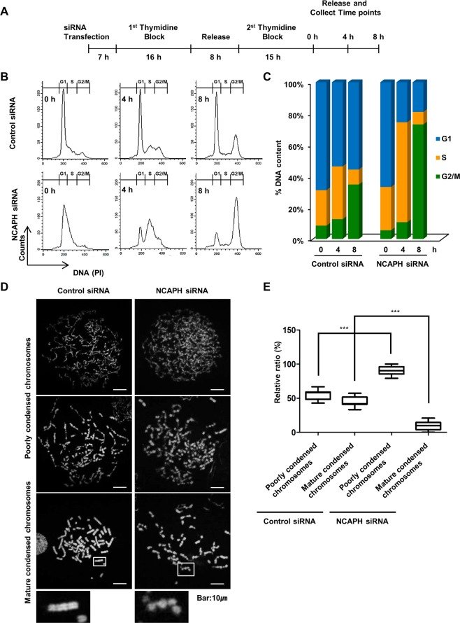
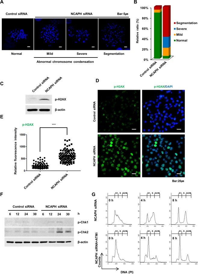
그림 출처: PubMed Central 공개 논문 (PMC6884527)
Conflict of interest statement
The authors declare no competing interests.
그 밖의 정보
- 출판 형태
- Journal Article, Research Support, Non-U.S. Gov't
- 저널
- Scientific reports · eISSN 2045-2322
- 전자 출판
- 2019-11-29
- MeSH
- Apoptosis · Caspase 3 / metabolism · Cell Cycle Checkpoints / drug effects · Cell Cycle Proteins / antagonists & inhibitors / genetics / metabolism · Cell Line, Tumor · Checkpoint Kinase 1 / metabolism · Checkpoint Kinase 2 / metabolism · Chromosome Aberrations* · DNA Damage* · Humans · Kaplan-Meier Estimate · Nuclear Proteins / antagonists & inhibitors / genetics / metabolism · Pancreatic Neoplasms / metabolism / mortality / pathology · RNA Interference · RNA, Small Interfering / metabolism
- Substances
- Cell Cycle Proteins · NCAPH protein, human · Nuclear Proteins · RNA, Small Interfering · Checkpoint Kinase 2 · Checkpoint Kinase 1 · Caspase 3
- 참고문헌
- 46편