
Ann Oncol. 2017;28(4):890-897.
Complex chromosomal rearrangements by single catastrophic pathogenesis in NUT midline carcinoma.
제1저자 이준구 · 교신저자 김태민, 주영석
소속 (7)
- Korea Advanced Institute of Science and Technology, Graduate School of Medical Science and Engineering, Daejeon, South Korea.
- Molecular Cytogenetics Core Facility, Wellcome Trust Sanger Institute, Wellcome Genome Campus, Hinxton, Cambridge, UK.
- Biomedical Science and Engineering Interdisciplinary Program, Korea Advanced Institute of Science and Technology, Daejeon, South Korea.
- Department of Laboratory Medicine, Chungnam National University School of Medicine, Daejeon, South Korea.
- Seoul National University Cancer Research Institute, Seoul, South Korea.
- Department of Internal Medicine Seoul National University Hospital, Seoul, South Korea.
- Pathology, Seoul National University Hospital, Seoul, South Korea.
Abstract
BACKGROUND Nuclear protein in testis (NUT) midline carcinoma (NMC) is a rare aggressive malignancy often occurring in the tissues of midline anatomical structures. Except for the pathognomonic BRD3/4-NUT rearrangement, the comprehensive landscape of genomic alterations in NMCs has been unexplored.
PATIENTS AND METHODS We investigated three NMC cases, including two newly diagnosed NMC patients in Seoul National University Hospital, and a previously reported cell line (Ty-82). Whole-genome and transcriptome sequencing were carried out for these cases, and findings were validated by multiplex fluorescence in situ hybridization and using individual fluorescence probes.
RESULTS Here, we present the first integrative analysis of whole-genome sequencing, transcriptome sequencing and cytogenetic characterization of NUT midline carcinomas. By whole-genome sequencing, we identified a remarkably similar pattern of highly complex genomic rearrangements (previously denominated as chromoplexy) involving the BRD3/4-NUT oncogenic rearrangements in two newly diagnosed NMC cases. Transcriptome sequencing revealed that these complex rearrangements were transcribed as very simple BRD3/4-NUT fusion transcripts. In Ty-82 cells, we also identified a complex genomic rearrangement involving the BRD4-NUT rearrangement underlying the simple t(15;19) karyotype. Careful inspections of rearrangement breakpoints indicated that these rearrangements were likely attributable to single catastrophic events. Although the NMC genomes had >3000 somatic point mutations, canonical oncogenes or tumor suppressor genes were rarely affected, indicating that they were largely passenger events. Mutational signature analysis showed predominant molecular clock-like signatures in all three cases (accounting for 54%-75% of all base substitutions), suggesting that NMCs may arise from actively proliferating normal cells.
CONCLUSION Taken together, our findings suggest that a single catastrophic event in proliferating normal cells could be sufficient for neoplastic transformation into NMCs.
© The Author 2017. Published by Oxford University Press on behalf of the European Society for Medical Oncology.
Keywords
NUT midline carcinomabromodomain and extra terminalchromoplexycomplex genomic rearrangementmutational signature
Figures
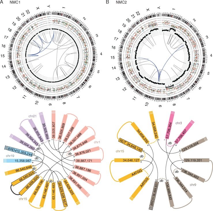
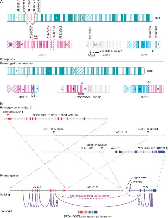
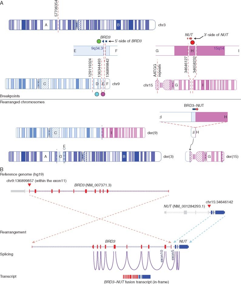
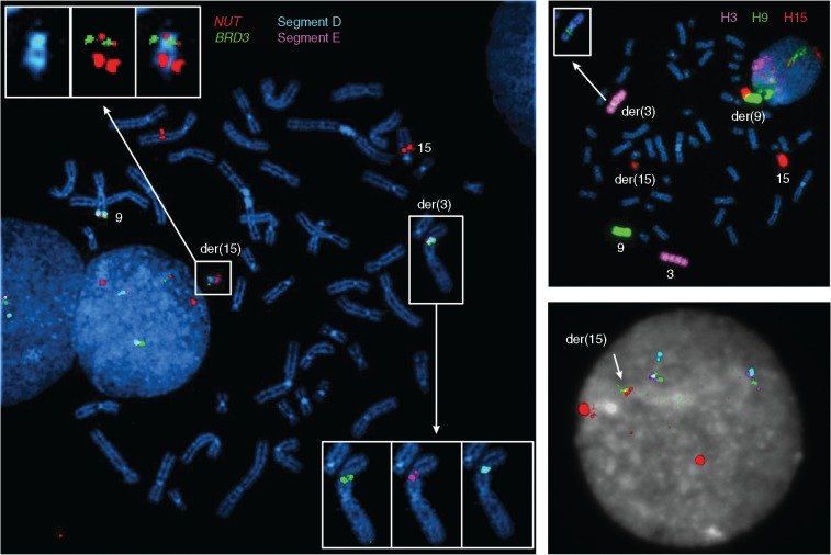
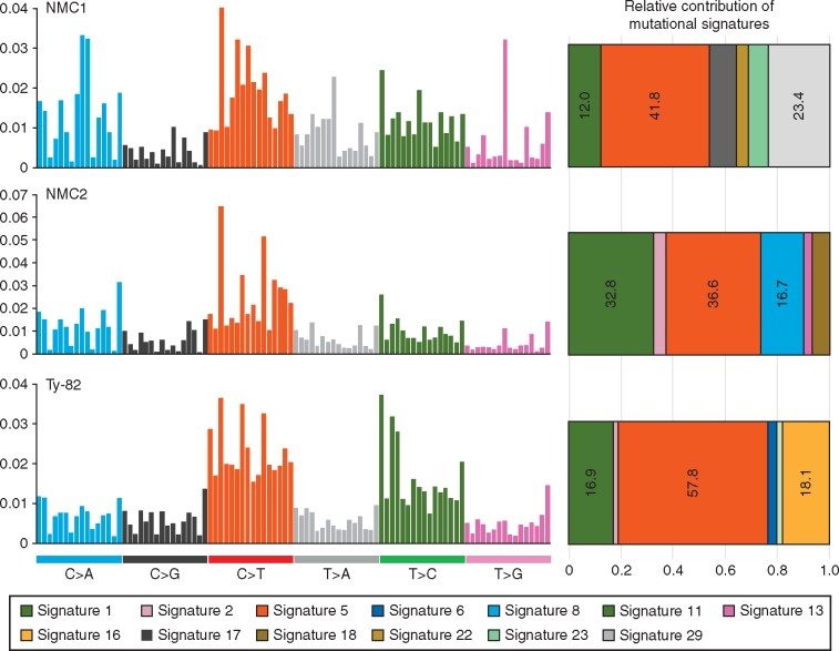
그림 출처: PubMed Central 공개 논문 (PMC5378225)
그 밖의 정보
- 출판 형태
- Case Reports, Journal Article
- 저널
- Annals of oncology : official journal of the European Society for Medical Oncology · eISSN 1569-8041
- MeSH
- Adult · Carcinoma / genetics · Cell Transformation, Neoplastic / genetics · Female · Gene Rearrangement · High-Throughput Nucleotide Sequencing · Humans · In Situ Hybridization, Fluorescence · Male · Nuclear Proteins / genetics · Oncogene Proteins, Fusion / genetics · RNA-Binding Proteins / genetics · Transcription Factors · Transcriptome · Bromodomain Containing Proteins
- Substances
- Nuclear Proteins · Oncogene Proteins, Fusion · RNA-Binding Proteins · Transcription Factors · BRD3 protein, human · Bromodomain Containing Proteins · BRD4-NUT fusion oncogene protein, human
- 참고문헌
- 24편