SCI 발표논문

SCI 발표논문

← SCI 발표논문 목록

Arthritis Res Ther. 2025;28(1):24.

Inhibition of NOX4 attenuates muscle damage and mitochondrial dysfunction in inflammatory myopathy.

, , , , , , ,

제1저자 강해림 · 교신저자 이은영

소속 (6)
  1. Interdisciplinary Program of Cancer Biology, Seoul National University College of Medicine, Seoul, Korea.
  2. Division of Rheumatology, Department of Internal Medicine, Seoul National University College of Medicine, Seoul, Korea.
  3. Inocras Korea, Inc., Daejeon, Republic of Korea.
  4. Interdisciplinary Program of Cancer Biology, Seoul National University College of Medicine, Seoul, Korea. elee@snu.ac.kr.
  5. Division of Rheumatology, Department of Internal Medicine, Seoul National University College of Medicine, Seoul, Korea. elee@snu.ac.kr.
  6. Division of Rheumatology, Department of Internal Medicine, Seoul National University Hospital, Seoul, Korea. elee@snu.ac.kr.

Abstract

BACKGROUND Idiopathic inflammatory myopathy (IIM) is a chronic autoimmune disorder characterized by muscle inflammation and weakness. If a muscle is already damaged, muscle strength often fails to restore with current treatments. Although, NADPH oxidase 4 (NOX4) is the main source of reactive oxygen species (ROS) and has been suggested to contribute to the pathogenesis of various disease by inducing ROS production and mitochondrial dysfunction, its role in IIM has not been fully explained.

METHODS Primary myoblasts from IIM patients and non-myopathic controls, as well as human skeletal muscle cells (SkMCs), were cultured under inflammatory conditions induced by interleukin (IL)-15, IL-6, and interferon-gamma (IFN-γ). NOX4 inhibitors, GKT137831 and GLX351322, were administered prior to cytokine stimulation. The effects of NOX4 inhibition were assessed in vitro and in vivo using a C protein-induced myositis (CIM) mouse model.

RESULTS IIM-derived myoblasts showed impaired myotube formation and elevated NOX4 expression compared to controls. Cytokine stimulation for SkMCs recapitulated key features of inflammatory myopathy, increased NOX4 and myoblast determination protein 1 (MyoD) expression. Treatment with NOX4 inhibitors reduced NOX4 and MyoD levels, restored myotube differentiation, and normalized the elevated oxygen consumption rate (OCR) associated with mitochondrial dysfunction. In the CIM model, NOX4 inhibition significantly reduced muscle inflammation (p < 0.05), preserved muscle mass (p < 0.001), increased cross-sectional area (CSA, p < 0.0001), and improved grip strength (p < 0.01).

CONCLUSIONS We showed that NOX4 is associated with muscle damage in IIM and suggest that its inhibition may have novel therapeutic effect for mitigating muscle damage and disease progression in IIM.

© 2025. The Author(s).

Keywords

InflammationInflammatory myopathyMitochondria dysfunctionMuscle regenerationMuscle weaknessNOX4

Figures
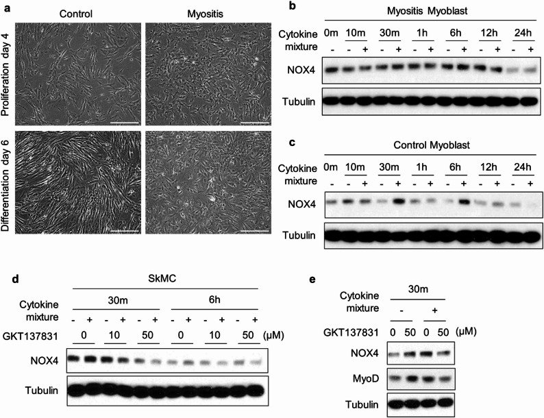
Fig. 1
Fig. 1 Differentiation defects and NOX4 overexpression in myoblasts from patients with IIM. a Myoblasts from controls and patients with IIM were cultured in 20% FBS and differentiated with 2% horse serum. Morphological differences between control and IIM myoblasts were observed at Day 4 of proliferation and Day 6 of differentiation. Morphology was observed by light microscopy at 500× magnification. b The expression of NOX4 in myoblasts from IIM patients was analyzed by immunoblotting after stimulation with either vehicle or a cytokine mixture (IL-15, IL-6, IFN-γ). Data represent six independent experiments (n = 6). c NOX4 protein expression in control myoblasts was assessed by immunoblotting after stimulation with either vehicle or the cytokine mixture (IL-15, IL-6, IFN-γ). Data represent three independent experiments (n = 3). d Skeletal muscle cells (SkMCs) were pretreated with 10 µM or 50 µM of GKT137831 for 4h, followed by stimulation with the cytokine mixture for 30 min, 6 h. NOX4 levels in SkMCs were analyzed by immunoblot. Data represent three independent experiments (n = 3). e NOX4 and MyoD levels were detected by immunoblot. Data represent three independent experiments (n = 3)
Fig. 2
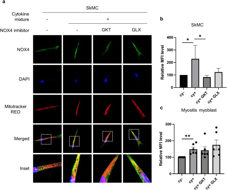
Fig. 2 Increased NOX4 localization in the mitochondria of inflammatory myoblasts. aSkMCs were pretreated with either 50 µM GKT137831 or 40 µM GLX351322 for 4h, followed by stimulation with vehicle or a cytokine mixture (IL-15, IL-6, and IFN-γ) for 30 min. Immunofluorescence staining was performed for NOX4 (green) and mitochondria (MitoTracker Red CMXRos, red), with nuclei were counterstained using DAPI (blue). Merged images show co-localization of NOX4 with mitochondria (yellow). Insets show 5x magnified views of regions marked by white boxes. b Mean fluorescence intensity (MFI) of mtROS was analyzed in SkMCs (n = 4) and c primary myoblasts derived from patients with IIM (n = 6), using FlowJo software. Data are presented as mean ± SEM
Fig. 3
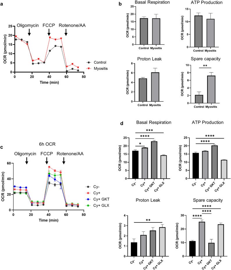
Fig. 3 Restoration of mitochondrial respiration abnormalities by inhibition of NOX4 in inflammatory myoblasts. aMitochondrial respiration was assessed using a Seahorse XFe96 Extracellular Flux Analyzer in primary myoblasts derived from a healthy control and a patient with idiopathic inflammatory myopathy. The oxygen consumption rate (OCR) was measured using the Mito Stress Test (n = 2). b Basal respiration, ATP production, proton leak, and spare respiratory capacity were calculated from OCR values. Group differences were analyzed using an unpaired t-test. c SkMCs were pretreated with 50 µM GKT137831 or 40 µM GLX351322 for 4h, followed by stimulation with either vehicle or a cytokine mixture (IL-15, IL-6, and IFN-γ) for 6h. d Mitochondrial function parameters –including basal respiration, ATP production, proton leak, and spare capacity- were quantified based on OCR. Group differences were assessed using one-way ANOVA (n = 3). Data are presented as mean ± SEM. *p < 0.05, **p < 0.01, ***p < 0.001, ****p < 0.0001
Fig. 4
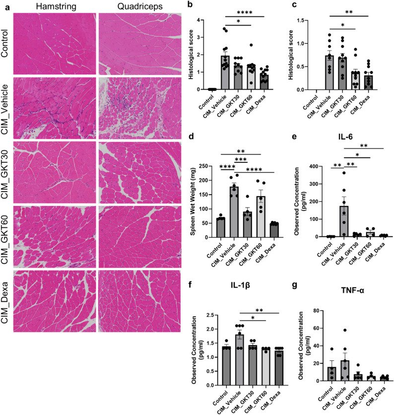
Fig. 4 NOX4 inhibition alleviates muscle inflammation in CIM model. aCIM was induced in C57BL/6 mice via immunization with C protein fragments, pertussis toxin, and complete Freund’s adjuvant. Mice received daily oral doses of GKT137831 (30 or 60 mg/kg) or dexamethasone from the day of immunization and were sacrificed on day 14. Muscle tissue sections were stained with H&E for histological analysis. b Inflammatory cell infiltration in muscle tissues after 14 days of immunization was graded on a scale from 0 to 6 (severe). c Inflammatory cell infiltration in muscle tissues 21 days after immunization was graded on a scale from 0 to 6. d Spleen weight was measured on day 14 to assess systemic inflammation. e, f Serum levels of pro-inflammatory cytokines IL-6, IL-1β and TNF-α were quantified using a multiplex immunoassay. Data are presented as mean ± SEM. Statistical significance was assessed using one-way ANOVA followed by post hoc testing. *p < 0.05, **p < 0.01, ***p < 0.001, ****p < 0.0001
Fig. 5
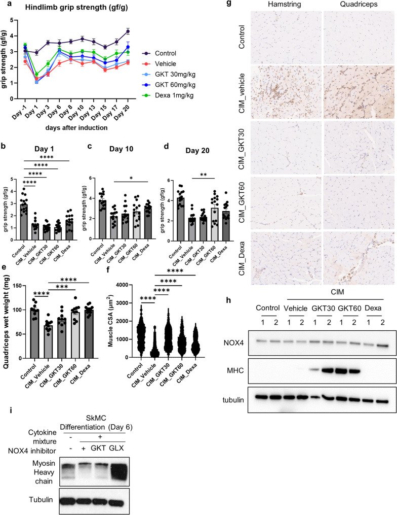
Fig. 5 Therapeutic potential of NOX4 inhibition in restoring muscle function and regeneration. aFemale C57BL/6 mice (7 weeks old) were injected with 200 µg C-protein emulsified in and sacrificed at day 14 or 21. Longitudinal assessment of hind limb grip strength until day 21. b-d Grip strength at days 1, 10, and 20. e Quantification of quadriceps muscle weight at sacrifice. f Cross-sectional area (CSA) of quadriceps muscle measured by ImageJ. g Representative NOX4-stained sections of quadriceps and hamstrings (200x magnification). h Immunoblot analysis of NOX4 and MHC expression in quadriceps muscle. i SkMCs were differentiated with 2% horse serum, treated with GKT137831 or GLX351322, and stimulated with a cytokine mixture (IL-15, IL-6, and IFN-γ) for 30 min. Data are presented as mean ± SEM. Statistical significance was assessed using one-way ANOVA followed by post hoc testing. *p < 0.05, **p < 0.01, ***p < 0.001, ****p < 0.0001

그림 출처: PubMed Central 공개 논문 (PMC12860102)

Conflict of interest statement

Declarations. Ethics approval and consent to participate: This study was approved by the Institutional Review Board (IRB) of Seoul National University Hospital (IRB No. 1902-120-1013). Informed consent was obtained from all patients. All animal procedures were approved by the Institutional Animal Care and Use Committee (IACUC) of Seoul National University Hospital (IACUC No. SNU-220727-1-2), and the experiments were conducted in accordance with institutional guidelines for the care and use of animals. Consent for publication: Not applicable. Competing interests: The authors declare no competing interests.

그 밖의 정보
출판 형태
Journal Article, Research Support, Non-U.S. Gov't
저널
Arthritis research & therapy · eISSN 1478-6362
전자 출판
2025-12-08
MeSH
NADPH Oxidase 4* / antagonists & inhibitors / metabolism · Humans · Animals · Mice · Myositis* / pathology / metabolism / drug therapy · Muscle, Skeletal* / drug effects / pathology / metabolism · Male · Female · Cells, Cultured · Mitochondria* / metabolism / drug effects · Reactive Oxygen Species / metabolism · Mice, Inbred C57BL · Middle Aged · Myoblasts / drug effects / metabolism · Benzoxazoles · Triazoles
Substances
NADPH Oxidase 4 · NOX4 protein, human · Reactive Oxygen Species · 3-benzyl-7-(2-benzoxazolyl)thio-1,2,3-triazolo(4,5-d)pyrimidine · Benzoxazoles · Triazoles
참고문헌
37편