
Gastric Cancer. 2024;27(5):1088-1099.
An artificial intelligence system for comprehensive pathologic outcome prediction in early gastric cancer through endoscopic image analysis (with video).
제1저자 이승한 · 교신저자 조수정
소속 (7)
- Department of Internal Medicine and Liver Research Institute, Seoul National University College of Medicine, 101 Daehak-Ro, Jongno-Gu, Seoul, 03080, Republic of Korea.
- Ainex Corporation, Seoul, Republic of Korea.
- Department of Internal Medicine, Seoul National University Bundang Hospital, Seoungnam-Si, Gyeonggi-Do, Republic of Korea.
- Center for Health Promotion and Optimal Aging, Seoul National University Hospital, Seoul, Republic of Korea.
- Department of Surgery, Seoul National University College of Medicine, Seoul, Republic of Korea.
- Department of Pathology, Seoul National University College of Medicine, Seoul, Republic of Korea.
- Department of Internal Medicine and Liver Research Institute, Seoul National University College of Medicine, 101 Daehak-Ro, Jongno-Gu, Seoul, 03080, Republic of Korea. crystal5@snu.ac.kr.
Abstract
BACKGROUND Accurate prediction of pathologic results for early gastric cancer (EGC) based on endoscopic findings is essential in deciding between endoscopic and surgical resection. This study aimed to develop an artificial intelligence (AI) model to assess comprehensive pathologic characteristics of EGC using white-light endoscopic images and videos.
METHODS To train the model, we retrospectively collected 4,336 images and prospectively included 153 videos from patients with EGC who underwent endoscopic or surgical resection. The performance of the model was tested and compared to that of 16 endoscopists (nine experts and seven novices) using a mutually exclusive set of 260 images and 10 videos. Finally, we conducted external validation using 436 images and 89 videos from another institution.
RESULTS After training, the model achieved predictive accuracies of 89.7% for undifferentiated histology, 88.0% for submucosal invasion, 87.9% for lymphovascular invasion (LVI), and 92.7% for lymph node metastasis (LNM), using endoscopic videos. The area under the curve values of the model were 0.992 for undifferentiated histology, 0.902 for submucosal invasion, 0.706 for LVI, and 0.680 for LNM in the test. In addition, the model showed significantly higher accuracy than the experts in predicting undifferentiated histology (92.7% vs. 71.6%), submucosal invasion (87.3% vs. 72.6%), and LNM (87.7% vs. 72.3%). The external validation showed accuracies of 75.6% and 71.9% for undifferentiated histology and submucosal invasion, respectively.
CONCLUSIONS AI may assist endoscopists with high predictive performance for differentiation status and invasion depth of EGC. Further research is needed to improve the detection of LVI and LNM.
© 2024. The Author(s).
Keywords
Artificial intelligenceConvolutional neural networkEarly gastric cancerEndoscopy
Figures
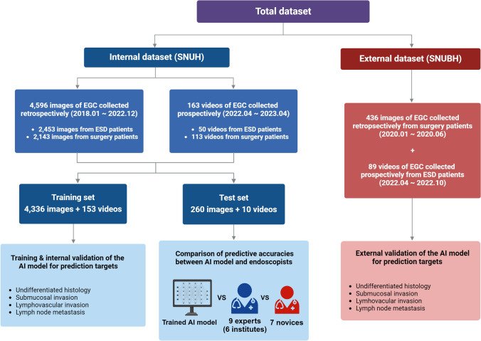
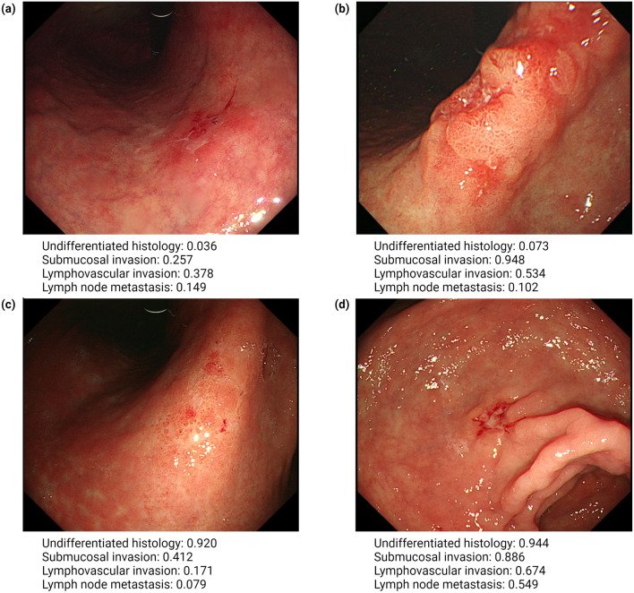
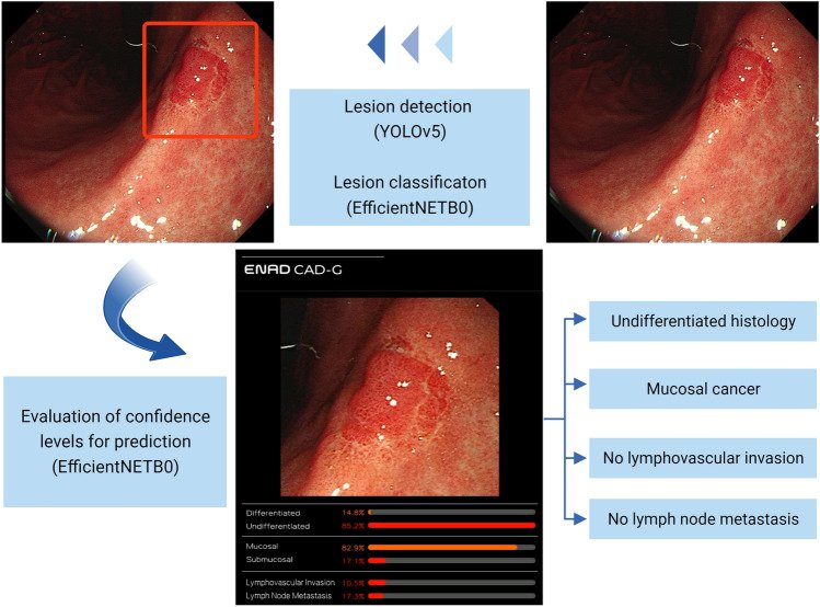
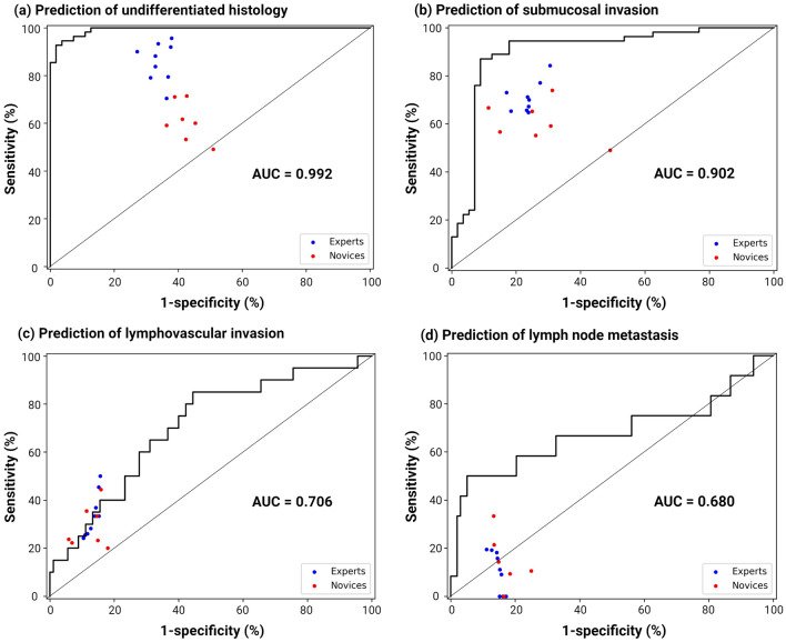
그림 출처: PubMed Central 공개 논문 (PMC11335909)
Comment · 정정
그 밖의 정보
- 출판 형태
- Journal Article, Research Support, Non-U.S. Gov't
- 저널
- Gastric cancer : official journal of the International Gastric Cancer Association and the Japanese Gastric Cancer Association · eISSN 1436-3305
- 전자 출판
- 2024-07-02
- MeSH
- Humans · Stomach Neoplasms* / pathology / surgery / diagnostic imaging · Artificial Intelligence* · Male · Female · Middle Aged · Retrospective Studies · Aged · Gastroscopy / methods · Prospective Studies · Image Processing, Computer-Assisted / methods · Early Detection of Cancer / methods · Adult · Lymphatic Metastasis / pathology · Aged, 80 and over · Neoplasm Invasiveness · Video Recording
- 연구비
- National Research Foundation of Korea #NRF-2022R1A2B5B01001430
- 참고문헌
- 47편