Department of Internal Medicine, Seoul National University Hospital, 101 Daekak-Ro, Chongno-Gu, Seoul, 03080, Korea.
National Research Laboratory for Stem Cell Niche, Seoul, Korea.
Innovative Research Institute for Cell Therapy, Seoul National University Hospital, Seoul, Korea.
Biometrology Group, Korea Research Institute of Standards and Science (KRISS), Daejeon, Korea.
Department of Internal Medicine, Soon Chun Hyang University Hospital, Bucheon, Korea.
Department of Internal Medicine, Seoul National University Hospital, 101 Daekak-Ro, Chongno-Gu, Seoul, 03080, Korea. hyosoo@snu.ac.kr.
National Research Laboratory for Stem Cell Niche, Seoul, Korea. hyosoo@snu.ac.kr.
Innovative Research Institute for Cell Therapy, Seoul National University Hospital, Seoul, Korea. hyosoo@snu.ac.kr.
Molecular Medicine and Biopharmaceutical Sciences, Seoul National University, Seoul, 03080, Korea. hyosoo@snu.ac.kr.
Abstract
BACKGROUND Although vasospastic angina (VSA) is known to be caused by coronary artery spasm, no study has fully elucidated the exact underlying mechanism. Moreover, in order to confirm VSA, patients should undergo invasive coronary angiography with spasm provocation test. Herein, we investigated the pathophysiology of VSA using peripheral blood-derived induced pluripotent stem cells (iPSCs) and developed an ex vivo diagnostic method for VSA.
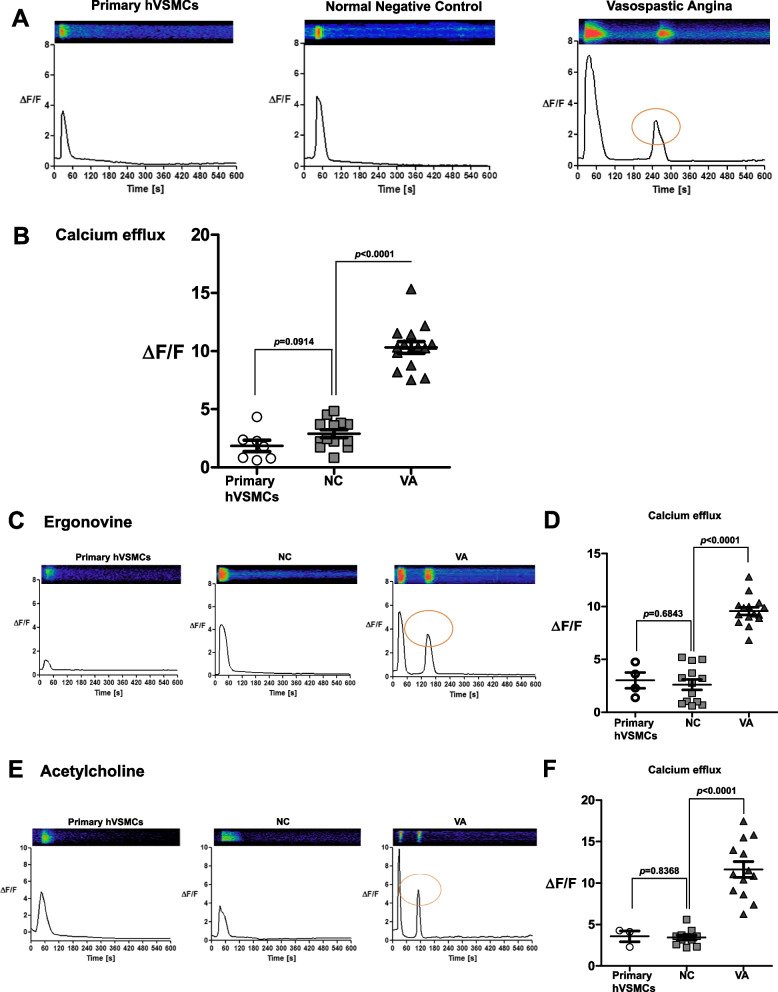
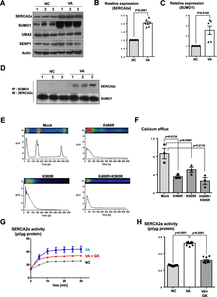
METHODS AND RESULTS With 10 mL of peripheral blood from patients with VSA, we generated iPSCs and differentiated these iPSCs into target cells. As compared with vascular smooth muscle cells (VSMCs) differentiated from iPSCs of normal subjects with negative provocation test, VSA patient-specific iPSCs-derived VSMCs showed very strong contraction in response to stimulants. Moreover, VSA patient-specific VSMCs exhibited a significant increase in stimulation-induced intracellular calcium efflux (Changes in the relative fluorescence unit [ΔF/F]; Control group vs. VSA group, 2.89 ± 0.34 vs. 10.32 ± 0.51, p < 0.01), and exclusively induced a secondary or tertiary peak of calcium efflux, suggesting that those findings could be diagnostic cut-off values for VSA. The observed hyperreactivity of VSA patient-specific VSMCs were caused by the upregulation of sarco/endoplasmic reticulum Ca2+-ATPase 2a (SERCA2a) due to its enhanced small ubiquitin-related modifier (SUMO)ylation. This increased activity of SERCA2a was reversed by treatment with ginkgolic acid, an inhibitor of SUMOylated E1 molecules (pi/µg protein; VSA group vs. VSA + ginkgolic acid, 52.36 ± 0.71 vs. 31.93 ± 1.13, p < 0.01).
CONCLUSIONS Our findings showed that abnormal calcium handling in sarco/endoplasmic reticulum could be induced by the enhanced SERCA2a activity in patients with VSA, leading to spasm. Such novel mechanisms of coronary artery spasm could be useful for drug development and diagnosis of VSA.
CalciumCoronary artery spasmEx vivo diagnosisVasospastic angina
Figures
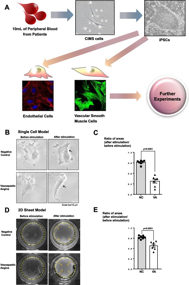
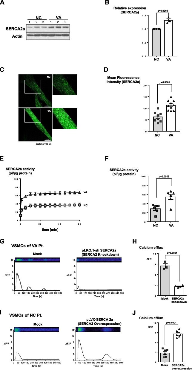
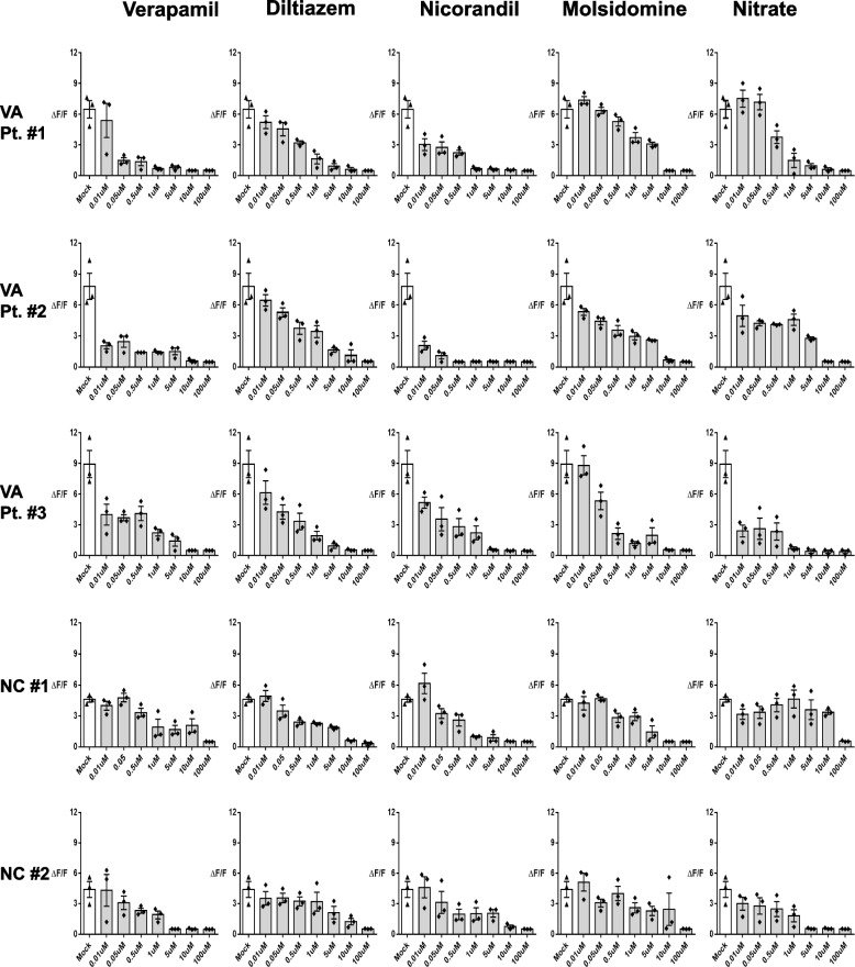
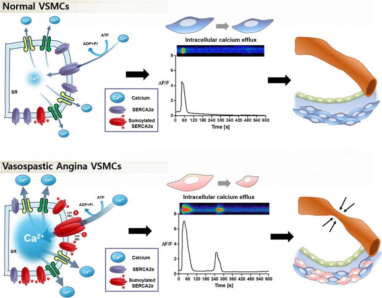
Fig. 1Hypercontractility of induced pluripotent stem cell (iPSC)-derived vascular smooth muscle cells (VSMCs) from vasospastic angina (VSA) patients. (A) Schematic summary of the study. We obtained 10 mL of peripheral blood from patients with VSA and cultured circulating multipotent stem (CiMS) cells. Thereafter, we generated VSA patient-specific iPSCs from these CiMS cells and differentiated iPSCs into both VSMCs and endothelial cells (ECs). (B, C) In a contraction assay using a single-cell model, 250 μM of carbachol treatment (a cholinomimetic vasoactive agonist) induced stronger contraction in VSMCs from VSA patients than in those from normal subjects. (n = 7 per group; scale bar, 5 μm). NC Negative control group with a negative provocation test, VA Vasospastic angina group with a positive provocation test. The black arrows denote the contraction area of the VSMCs. (D, E) Collagen gel contraction assays for 2 dimensional (2D) sheet models were examined using normal control subject-derived VSMCs and VSA patient-derived VSMCs. Treatment with 1 mM carbachol for 48 h induced a strong contraction of sheets derived from the VSMCs of VSA patients, as observed in the 2D sheet model (n = 7 per group). The areas circled with yellow dash lines denote the sizes of the collagen gels, and the yellow arrows indicate the contraction of the collagen gelsFig. 2Hyperreactivity of iPSC-derived VSMCs from VSA patients was triggered by an abnormal increase of intracellular calcium efflux (A, B) Intracellular calcium efflux in response to carbachol was measured with Fluo-4 (a calcium fluorescent dye. Fluo-4 treated cells were monitored through time-lapse confocal microscopy over 10 min after the addition of 250 μM of carbachol, an inducer of contraction. The iPSC-derived VSMCs from VSA patients exhibited much higher intensity of the calcium efflux peak than the cells from control subjects. Moreover, secondary or tertiary peaks of calcium efflux were observed only in the VSA group (primary human VSMCs n = 7; NC n = 13; VA n = 15). Primary human VSMCs indicate commercially available human coronary artery vascular smooth muscle cells. The orange circle indicates the secondary peak. (C-F) The addition of 250 μM ergonovine and 250 μM acetylcholine in primary human VSMCs, NC- and VSA- iPSC-derived VSMCs produced similar results to treated carbachol (Ergonovine; primary human VSMCs n = 4; NC n = 13; VA n = 15) (Acetylcholine; primary human VSMCs n = 3; NC n = 11; VA n = 13). The orange circle indicates the secondary peakFig. 3The mechanism of hyperreactivity of iPSC-derived VSMCs from VSA patient: the enhanced sarco/endoplasmic reticulum Ca2+-ATPase 2a (SERCA2a) activity. (A, B) 10 μg of whole protein lysates from each of the samples were loaded with a 10% SDS-PAGE gel, transferred to nitrocellulose membranes, and probed with anti-SERCA2a (1:1000) and anti-sarcomeric actin antibodies (1:1000). Western blot indicates that the amount of endogenous SERCA2a protein was greater in iPSC-derived VSMCs from VSA patients than from normal subjects (n = 3 per group). All samples in Western blot were obtained from individual subjects (three different normal subjects and three different VSA patients). (C, D) Immunofluorescence staining for SERCA2a shows higher intensity in iPSC-derived VSMCs from VSA patients than from normal subjects (n= 8–10 per group; scale bar, 20 μm). Each group of cells was stained with human anti-SERCA2a (1:100). SERCA2a was detected using the Alexa Fluor 488-conjugated anti-mouse secondary antibody (1:100). (E, F) Calcium-dependent ATPase activity assay indicates that the activity of SERCA2a in the VSA group was greater than in the control group (n = 6 per group). 10 μg of each cell lysate was mixed with the reaction buffer and pre-incubated for 10 min before being treated with 1 mM adenosine triphosphate (ATP). The ATPase activity was measured at 360 nm using a microplate reader. (G, H) After knocking down SERCA2a expression in iPSC-derived VSMCs from VSA patients using a lentiviral vector and induced with 250 μM carbachol, intracellular calcium efflux was downregulated to the levels of control cells (Mock n = 3; shSERCA2a n = 4). Mock indicates control lentiviral vector transfection. (I, J) In contrast, the overexpression of SERCA2a in iPSC-derived VSMCs from normal subjects using a lentiviral vector induced with 250 μM carbachol increased the level of intracellular calcium efflux and generated the second peak of calcium efflux (n = 6 per group)Fig. 4The enhanced SERCA2a activity of iPSC-derived VSMCs from VSA patient was induced by the increased small ubiquitin-related modifier (SUMO)ylation of SERCA2a. (A-C) Western blot shows that the amount of endogenous SERCA2a and SUMO1 proteins was greater in VSA patient-derived VSMCs than in normal subject-derived VSMCs (n = 4–7 per group). 50 μg of whole protein lysates from each sample were loaded with a 10% SDS-PAGE gel, transferred to nitrocellulose membranes, and probed with anti-SERCA2a (1:1000), anti-SUMO1 (1:1000), anti-UBA2 (1:1000), anti-SENP1 (1:1000), and anti-sarcomeric actin antibodies (1:1000). SUMO1 = Small Ubiquitin Like Modifier 1; UBA2 = Ubiquitin-like modifier-activating enzyme 2; SENP1 = Sentrin-specific protease 1. (D) Immunoprecipitation assay for SUMO1 and SERCA2a indicates that SUMO1 bound only to SERCA2a of VSMCs from VSA patients, not from normal subjects. (E, F) We transfected VSA-patient derived VSMCs with lentiviral vectors encoding SERCA2a with mutated lysine 480 and 585 residues. Each group was treated with Fluo-4 and monitored through time-lapse confocal microscopy over 10 min after the addition of 250 μM carbachol. The transfection with the mutant forms exhibited lower intracellular calcium efflux, indicating that these SUMOylated sites are responsible for the increased activity of SECAR2a in the VSMCs from VSA patients (n = 3 per group). (G, H) 10 μg of each cell lysate was mixed with the reaction buffer and treated with 1 mM ATP and Ginkgolic acid (10 μM), an inhibitor of SUMOylated E1 molecules. Ginkgolic acid suppressed enhanced SERCA2a activity in iPSC-derived VSMCs from VSA patients (n = 9 per group). GK Ginkgolic acidFig. 5Differences in the magnitude of intracellular calcium efflux among VSA patients in response to various dosages of diverse vasodilators. By measuring intracellular calcium efflux in iPSC-derived VSMCs from VSA patients or normal subjects via our method, we evaluated changes of intracellular calcium efflux in response to various dosages (0.01, 0.05, 0.5, 1, 5, 10, 100 μM) of calcium channel blockers (Verapamil, Diltiazem) and vasodilators (Nicorandil, Molsidomine, Nitrate). The inhibitory effect of each drug differed even among VSA patientsFig. 6Schematic figure of the mechanism underlying VSMCs hyper-responsiveness in VSA patients. Compared to the status in normal subjects, the increased SUMOylation of SERCA2a in VSA patients enhances its stability, amount, and activity, in turn increasing intracellular calcium efflux and VSMCs hyperreactivity leading to vessel contraction
그림 출처: PubMed Central 공개 논문 (PMC9938986)
Conflict of interest statement
None of the authors have any financial or personal interests to disclose.
그 밖의 정보
출판 형태
Journal Article
저널
Biomaterials research · ISSN 1226-4601
전자 출판
2023-02-18
연구비
Korea Health Industry Development Institute HI14C1277 · Korea Health Industry Development Institute HI17C2085 · Ministry of Education, Science and Technology 2020R1A2C1011311