SCI 발표논문

SCI 발표논문

← SCI 발표논문 목록

Clin Mol Hepatol. 2020;26(4):529-539.

Inhibition of PI3K/Akt signaling suppresses epithelial-to-mesenchymal transition in hepatocellular carcinoma through the Snail/GSK-3/beta-catenin pathway.

, , , , , , ,

제1저자 이슬기, 최은지 · 교신저자 김윤준

소속 (1)
  1. Department of Internal Medicine, Seoul National University College of Medicine, Seoul, Korea.

Abstract

BACKGROUND/AIMS Patients with advanced hepatocellular carcinoma (HCC) have a poor prognosis due to the lack of effective systemic therapies. Epithelial-to-mesenchymal transition (EMT) is a pivotal event in tumor progression, during which cancer cells acquire invasive properties. In this study, we investigated the effects of phosphatidylinositol 3-kinase (PI3K) inhibitors, including LY294002 and idelalisib, on the EMT features of HCC cells in vitro.

METHODS Human HCC cell lines, including Huh-BAT and HepG2, were used in this study. Cell proliferation was measured by 3-(4,5-dimethylthiazol-2-yl)-2,5-diphenyl tetrazolium bromide, and cell cycle distributions were evaluated using a flow cytometer by propidium iodide staining. Immunofluorescence staining, quantitative real-time polymerase chain reaction, and immunoblotting were performed to detect EMT-associated changes.

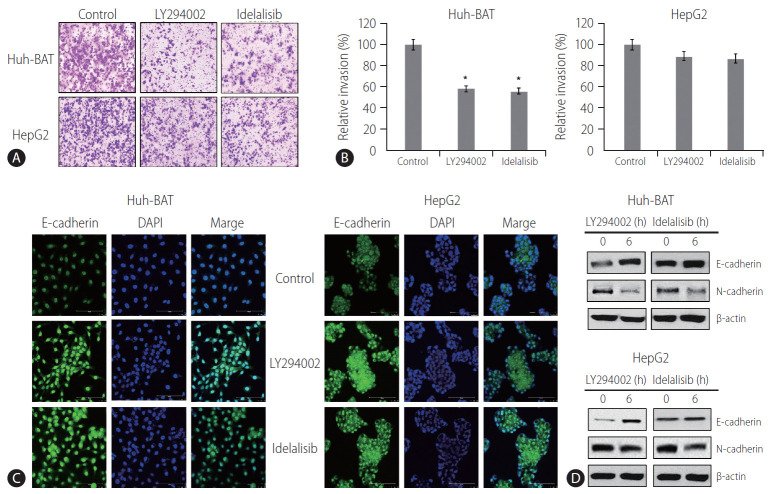
RESULTS PI3K inhibitors suppressed the proliferation and invasion of HCC cells and deregulated the expression of EMT markers, as indicated by increased expression of E-cadherin, an epithelial marker, and decreased expression of N-cadherin, a mesenchymal marker, and Snail, a transcription factor implicated in EMT regulation. Furthermore, LY294002 and idelalisib inhibited the phosphorylation of GSK-3β and induced the nuclear translocation of GSK-3β, which corresponded to the downregulation of Snail and β-catenin expressions in Huh-BAT and HepG2 cells.

CONCLUSION The inhibition of PI3K/Akt signaling decreases Snail expression by enhancing the nuclear translocation of GSK-3β, which suppresses EMT in HCC cells, suggesting the potential clinical application of PI3K inhibitors for HCC treatment.

Keywords

Carcinoma, HepatocellularEpithelial-to-mesenchymal transitionGlycogen synthase kinase-3Phosphatidylinositol 3-kinasesSnails

Figures

Figure 1.
Figure 1. LY294002 and idelalisib reduce cellular proliferation and induce cell cycle arrest at the G0/G1 phase. (A) Huh-BAT and HepG2 cells were treated with 20 μM LY294002 or idelalisib for 48 hours. Cell morphology was observed by phase-contrast microscopy. Scale bar, 100 μm. (B) Huh-BAT and HepG2 cells were treated with different concentrations of LY294002 or idelalisib (5, 10, and 20 μM) for 2, 6, 24, and 48 hours. Cell viability was measured using the MTT assay. Flow cytometry analysis were monitored to measure cell cycle distributions (C, D). Cells were treated with 20 μM LY294002 or idelalisib for 48 hours. The percentage of cell cycle progression was evaluated from three independent experiments. (E) The protein expression of cyclin D, cyclin E and CDK6, total Akt, and phosphor-Akt was detected by immunoblotting, and quantifications are shown. Data are presented as the mean±standard deviation. *P<0.05 and †P<0.01 compared with untreated control cells.
Figure 2.
Figure 2. LY294002 and idelalisib suppress the epithelial-mesenchymal transition. (A, B) Huh-BAT and HepG2 cells were treated with 20 μM LY294002 or idelalisib, for 48 hours, and invasion capability was measured with an invasion assay. The resulting colonies were stained and quantified. All data are expressed as the mean±standard deviation. *P<0.05. (C) Huh-BAT and HepG2 cells were treated with 20 μM LY294002 or idelalisib, for 48 hours. E-cadherin expression was measured by confocal fluorescence microscopy (magnification, ×400). (D) Changes in the protein levels of E-cadherin and N-cadherin were analyzed by immunoblotting following 48 hours of treatment with 20 μM LY294002 or idelalisib. DAPI, 4',6-diamidino-2-phenylindole.
Figure 3.
Figure 3. LY294002 and idelalisib suppress Snail and β-catenin expressions. (A, B) The mRNA levels of Snail and β-catenin were quantified by qRT-PCR following treatment with 20 μM LY294002 or idelalisib in Huh-BAT and HepG2 cells. (C, D) Huh-BAT cells and HepG2 cells were treated with 20 μM LY294002 or idelalisib, for various time points. The protein expression levels of Snail were evaluated by immunoblotting. β-actin was used as a loading control. (E, F) Huh-BAT cells and HepG2 cells were treated with 20 μM LY294002 or idelalisib, for various time points. The protein expression levels of Snail and phospho-GSK-3β were evaluated by immunoblotting. β-actin was used as a loading control. Data are presented as the mean±standard deviation. *P<0.05 and †P<0.01 compared with untreated control cells. P-GSK, phospho-glycogen synthase kinase; qRT-PCR, quantitative Real-time polymerase chain reaction.
Figure 4.
Figure 4. LY294002 and idelalisib regulate cellular translocalization of proteins. (A) Huh-BAT cells were treated with 20 μM LY294002 or idelalisib for 48 hours. Protein expression levels of β-catenin, GSK-3β and Snail were evaluated by immunoblotting in both nuclear and cytosolic compartments. α-tubulin and HDAC1 were used as nuclear and cytosolic loading controls, respectively. (B) Huh-BAT cells were treated with 20 μM LY294002 or idelalisib for 48 hours. Following treatments, localization of GSK-3β was evaluated using immunofluorescence. GSK-3β, glycogen synthase kinase-3β; DAPI, 4',6-diamidino-2-phenylindole.

그림 출처: PubMed Central 공개 논문 (PMC7641573)

Comment · 정정

Conflict of interest statement

Conflicts of Interest: The authors have no conflicts to disclose.

그 밖의 정보
출판 형태
Journal Article, Research Support, Non-U.S. Gov't
저널
Clinical and molecular hepatology · eISSN 2287-285X
전자 출판
2020-08-24
MeSH
Carcinoma, Hepatocellular / therapy · Catenins · Cell Line, Tumor · Glycogen Synthase Kinase 3 · Glycogen Synthase Kinase 3 beta · Humans · Liver Neoplasms* · Phosphatidylinositol 3-Kinases · Proto-Oncogene Proteins c-akt · beta Catenin
Substances
Catenins · Glycogen Synthase Kinase 3 · Glycogen Synthase Kinase 3 beta · Phosphatidylinositol 3-Kinases · Proto-Oncogene Proteins c-akt · beta Catenin
참고문헌
37편