
PLoS One. 2014;9(6):e98232.
The effect of lithospermic acid, an antioxidant, on development of diabetic retinopathy in spontaneously obese diabetic rats.
제1저자 Jin CJ · 교신저자 임수
소속 (8)
- Department of Internal Medicine, Seoul National University College of Medicine and Seoul National University Bundang Hospital, Seongnam, Korea; Department of Endocrinology, The ZhongShan Hospital, DaLian University, Dalian, China.
- Department of Internal Medicine, Seoul National University College of Medicine and Seoul National University Bundang Hospital, Seongnam, Korea; Department of Internal Medicine, Hallym University Hangang Sacred Hospital, Seoul, Korea.
- Department of Endocrinology, The ZhongShan Hospital, DaLian University, Dalian, China.
- Department of Ophthalmology, Seoul National University College of Medicine and Seoul National University Bundang Hospital, Seongnam, Korea.
- Department of Pathology, Seoul National University College of Medicine and Seoul National University Bundang Hospital, Seongnam, Korea.
- Department of Internal Medicine, Yonsei University College of Medicine, Seoul, Korea.
- Department of Internal Medicine, Seoul National University College of Medicine and Seoul National University Bundang Hospital, Seongnam, Korea.
- Department of Internal Medicine, Seoul National University College of Medicine, Seoul, Korea.
Abstract
BACKGROUND Lithospermic acid B (LAB), an active component isolated from Salvia miltiorrhiza radix, has been reported to have antioxidant effects. We examined the effects of LAB on the prevention of diabetic retinopathy in Otsuka Long-Evans Tokushima Fatty (OLETF) rats, an animal model of type 2 diabetes.
METHODS AND FINDINGS LAB (10 or 20 mg/kg) or normal saline were given orally once daily to 24-week-old male OLETF rats for 52 weeks. At the end of treatment, fundoscopic findings, vascular endothelial growth factor (VEGF) expression in the eyeball, VEGF levels in the ocular fluid, and any structural abnormalities in the retina were assessed. Glucose metabolism, serum levels of high-sensitivity C-reactive protein (hsCRP), monocyte chemotactic protein-1 (MCP1), and tumor necrosis factor-alpha (TNFα) and urinary 8-hydroxy-2'-deoxyguanosine (8-OHdG) levels were also measured. Treatment with LAB prevented vascular leakage and basement membrane thickening in retinal capillaries in a dose-dependent manner. Insulin resistance and glucose intolerance were significantly improved by LAB treatment. The levels of serum hsCRP, MCP1, TNFα, and urinary 8-OHdG were lower in the LAB-treated OLETF rats than in the controls.
CONCLUSIONS Treatment with LAB had a preventive effect on the development of diabetic retinopathy in this animal model, probably because of its antioxidative effects and anti-inflammatory effects.
Figures
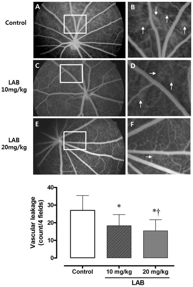
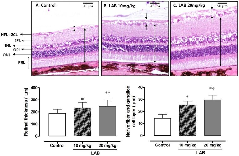
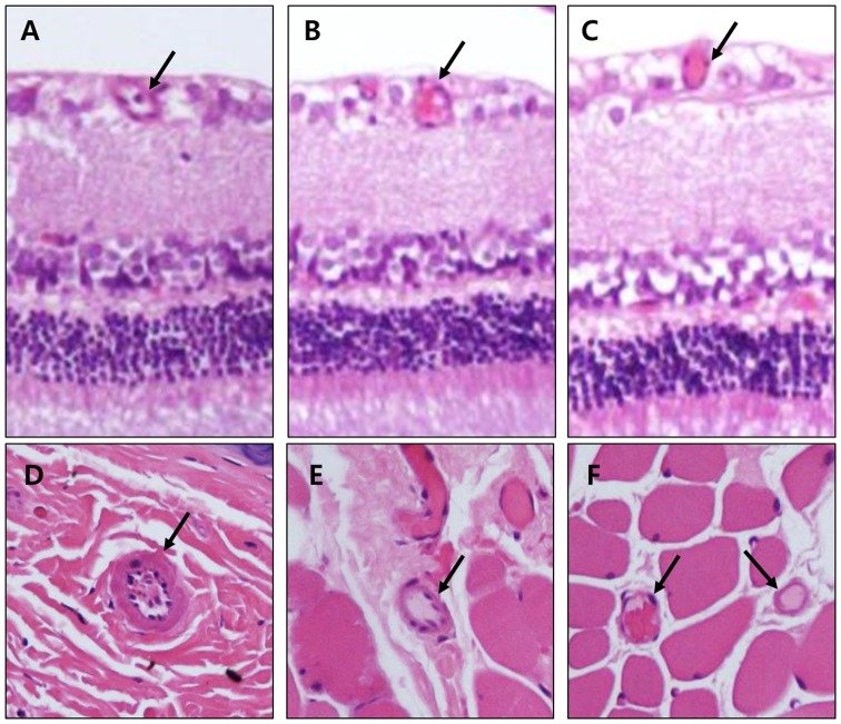
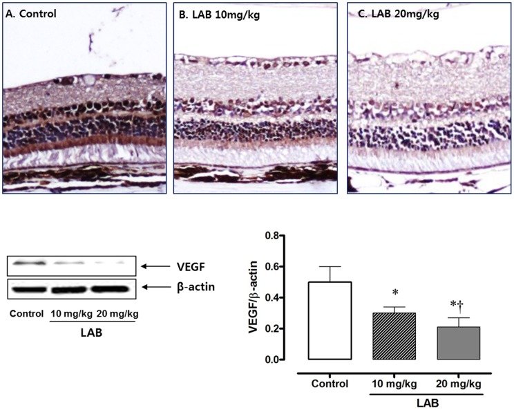
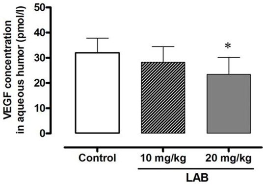
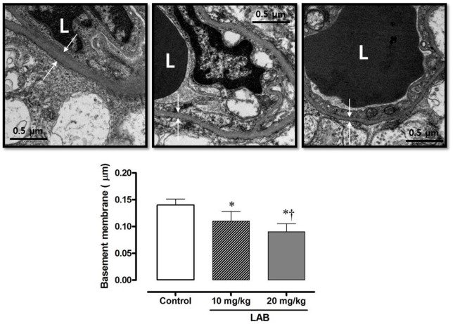
그림 출처: PubMed Central 공개 논문 (PMC4048190)
Conflict of interest statement
Competing Interests:The authors have declared that no competing interests exist.
그 밖의 정보
- 출판 형태
- Journal Article, Research Support, Non-U.S. Gov't
- 저널
- PloS one · eISSN 1932-6203
- 전자 출판
- 2014-06-06
- MeSH
- 8-Hydroxy-2'-Deoxyguanosine · Animals · Antioxidants / therapeutic use · Benzofurans / therapeutic use · C-Reactive Protein / metabolism · Chemokine CCL2 / metabolism · Deoxyguanosine / analogs & derivatives / urine · Depsides / therapeutic use · Diabetic Retinopathy / metabolism / prevention & control · Glucose / metabolism · Glucose Intolerance / drug therapy · Insulin Resistance · Male · Obesity / complications · Rats · Rats, Long-Evans · Tumor Necrosis Factor-alpha / metabolism · Vascular Endothelial Growth Factor A / metabolism
- Substances
- Antioxidants · Benzofurans · Chemokine CCL2 · Depsides · Tumor Necrosis Factor-alpha · Vascular Endothelial Growth Factor A · lithospermic acid · 8-Hydroxy-2'-Deoxyguanosine · C-Reactive Protein · Deoxyguanosine · Glucose
- 참고문헌
- 56편