
Sci Rep. 2019;9(1):4831.
Anatomic Conformation of Renal Sympathetic Nerve Fibers in Living Human Tissues.
제1저자 최원석 · 교신저자 최의근
소속 (5)
- Department of Internal Medicine, Seoul National University Hospital, Seoul, Republic of Korea.
- Department of Urology, Pusan National University Yangsan Hospital, Yangsan, Republic of Korea.
- Department of Urology, Seoul National University Hospital, Seoul, Republic of Korea.
- Department of Urology, Seoul National University Hospital, Seoul, Republic of Korea. drboss@korea.com.
- Department of Internal Medicine, Seoul National University Hospital, Seoul, Republic of Korea. choiek17@snu.ac.kr.
Abstract
Renal denervation using radiofrequency catheter ablation is known to eliminate the renal sympathetic nerve and to lower blood pressure in patients with resistant hypertension. We sought to investigate the detailed anatomic conformation of the peri-renal arterial sympathetic nerve fibers with living human specimens. Peri-renal arterial tissue was harvested from patients undergoing elective radical or simple nephrectomy. Digital images of each section from the distal arterial bifurcation to the proximal margin were obtained and analyzed after immunohistochemical staining with anti-tyrosine hydroxylase antibodies. A total of 3,075 nerve fibers were identified from 84 sections of peri-renal arterial tissue from 28 patients (mean age 62.5 ± 10.2 years, male 68%). Overall, 16% of nerve fibers were located at distances greater than 3 mm from the endoluminal surface of the renal artery. The median distance from the arterial lumen to the nerve fibers of the proximal, middle, and distal renal arterial segments was 1.51 mm, 1.48 mm, and 1.52 mm, respectively. The median diameter of the nerve fibers was 65 μm, and there was no significant difference between the segments. A substantial proportion of the sympathetic nerve fibers were located deeper in the peri-arterial soft tissue than in the lesion depth created by the conventional catheter-based renal sympathetic denervation system.
Figures
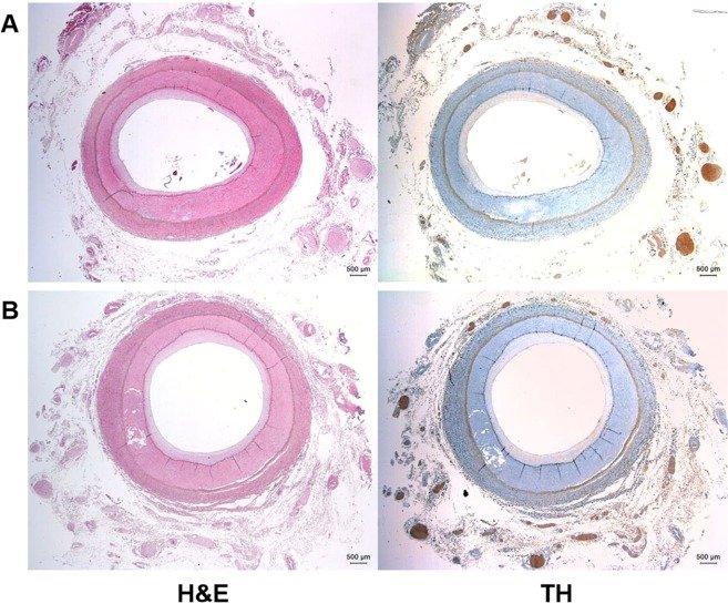
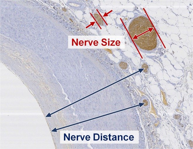
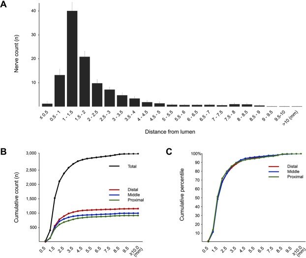
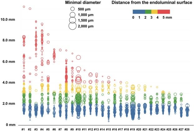
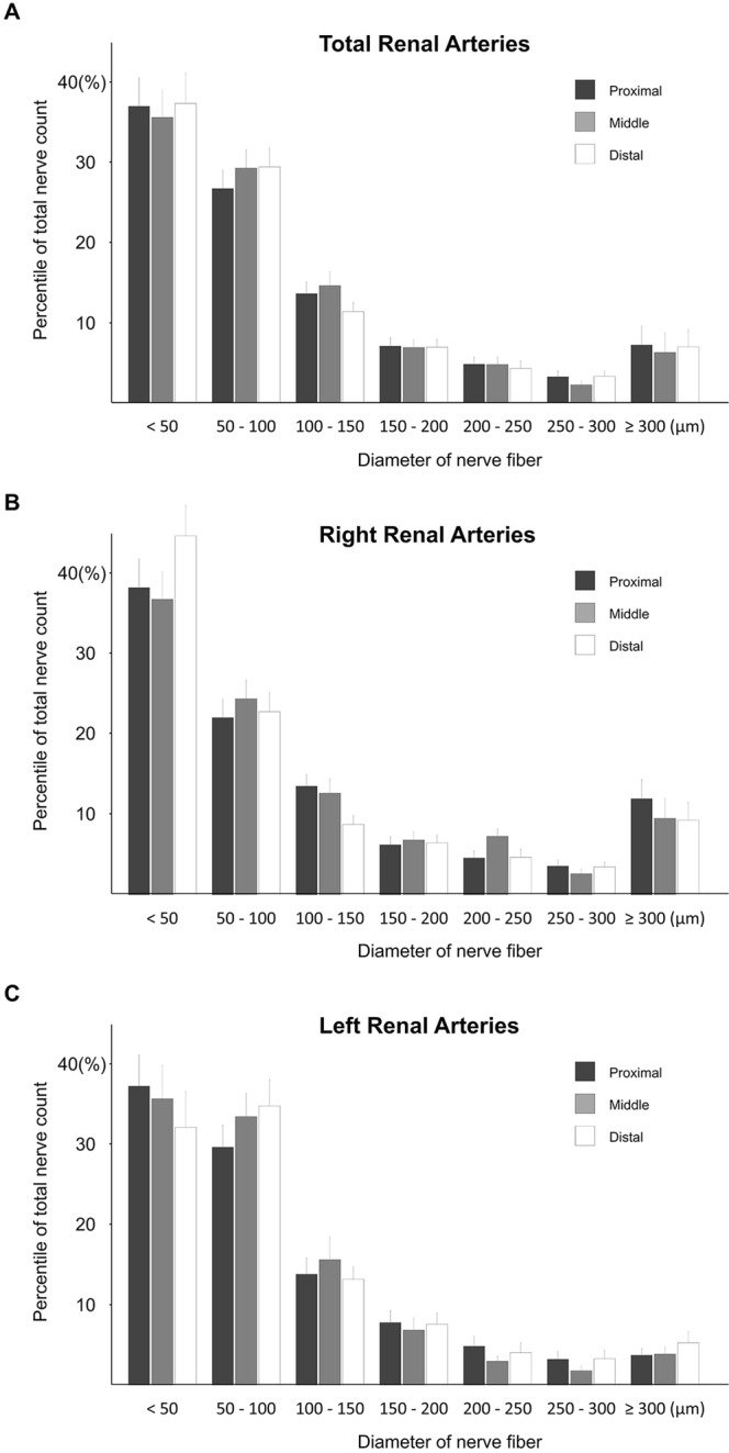
그림 출처: PubMed Central 공개 논문 (PMC6423056)
Conflict of interest statement
The authors declare no competing interests.
그 밖의 정보
- 출판 형태
- Journal Article, Observational Study, Research Support, Non-U.S. Gov't
- 저널
- Scientific reports · eISSN 2045-2322
- 전자 출판
- 2019-03-18
- MeSH
- Adrenergic Fibers / physiology · Aged · Antihypertensive Agents / pharmacology / therapeutic use · Blood Pressure / physiology · Catheter Ablation* · Drug Resistance · Female · Humans · Hypertension / physiopathology / therapy · Kidney / blood supply / innervation / physiology / surgery · Male · Middle Aged · Nephrectomy · Renal Artery / diagnostic imaging / innervation / physiology · Sympathectomy · Sympathetic Nervous System / anatomy & histology / diagnostic imaging / physiology
- Substances
- Antihypertensive Agents
- 참고문헌
- 38편