Biomedical Research Institute, Seoul National University Hospital, Seoul, 03080, Republic of Korea.
Department of Molecular Medicine and Biopharmaceutical Sciences, Graduate School of Convergence Science and Technology, Seoul National University, Seoul, 03080, Republic of Korea.
Department of Internal Medicine, Seoul National University College of Medicine, Seoul, 03080, Republic of Korea.
Department of Biomedical Sciences, Seoul National University College of Medicine, Seoul, 03080, Republic of Korea.
Department of Life Sciences, Pohang University of Science and Technology, Pohang, Gyeongbuk, 37673, Republic of Korea.
Biomedical Research Institute, Seoul National University Hospital, Seoul, 03080, Republic of Korea. ehakmo@gmail.com.
Veterans Medical Research Institute, Veterans Health Service Medical Center, Seoul, 05368, Republic of Korea. ehakmo@gmail.com.
Biomedical Research Institute, Seoul National University Hospital, Seoul, 03080, Republic of Korea. kspark@snu.ac.kr.
Department of Molecular Medicine and Biopharmaceutical Sciences, Graduate School of Convergence Science and Technology, Seoul National University, Seoul, 03080, Republic of Korea. kspark@snu.ac.kr.
Department of Internal Medicine, Seoul National University College of Medicine, Seoul, 03080, Republic of Korea. kspark@snu.ac.kr.
Abstract
Transplantation of stem cell-derived insulin producing cells (IPCs) has been proposed as an alternative to islet transplantation for the treatment of diabetes mellitus. However, current IPC differentiation protocols are focused on generating functional cells from the pluripotent stem cells and tend to rely on multistep, long-term exposure to various exogenous factors. In this study, we addressed the observation that under stress, pancreatic β-cells release essential components that direct the differentiation of the bone marrow nucleated cells (BMNCs) into IPCs. Without any supplementation with known differentiation-inducing factors, IPCs can be generated from BMNCs by in vitro priming for 6 days with conditioned media (CM) from the β-cells. In vitro primed BMNCs expressed the β-cell-specific transcription factors, as well as insulin, and improved hyperglycemia and glucose intolerance after transplantation into the streptozotocin-induced diabetic mice. Furthermore, we have found that components of the CM which trigger the differentiation were enclosed by or integrated into micro particles (MPs), rather than being secreted as soluble factors. Identification of these differentiation-directing factors might enable us to develop novel technologies required for the production of clinically applicable IPCs.
Figures
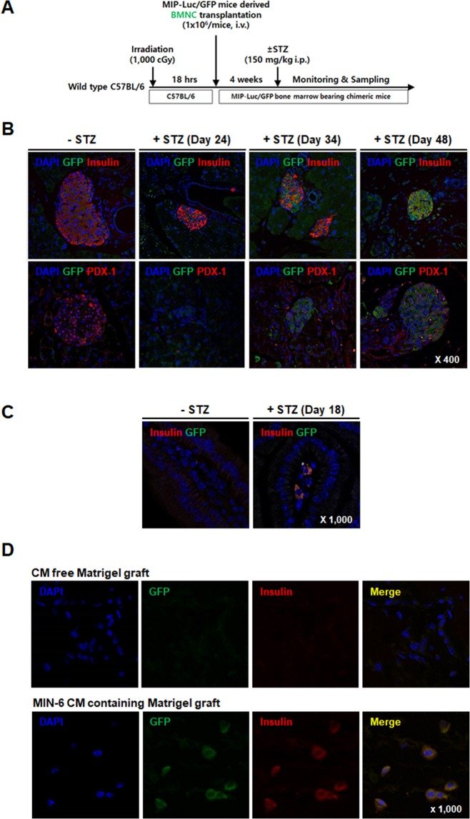
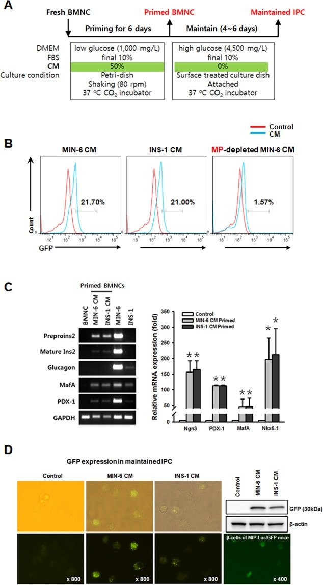
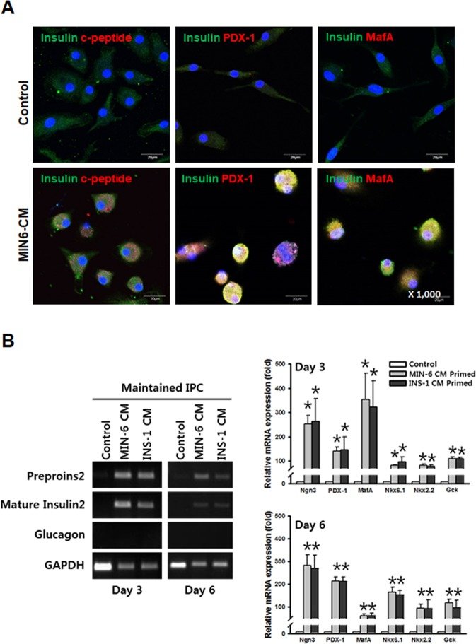
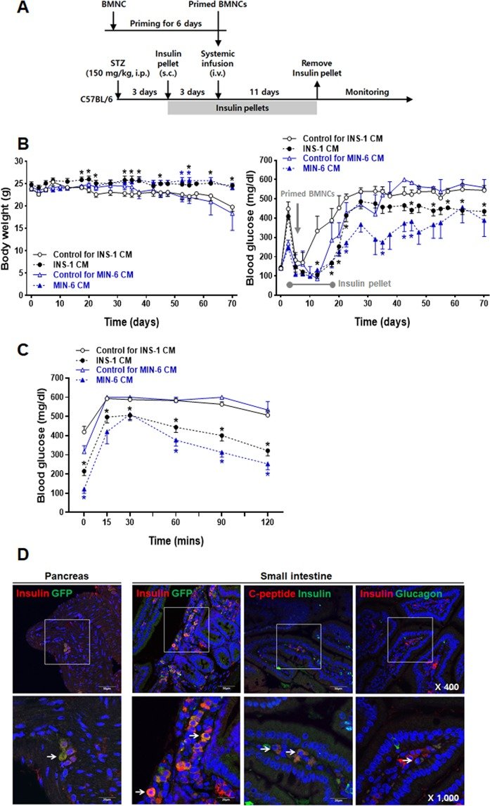
Figure 1BMNCs differentiate into IPCs in vivo. (A) Outline of the experimental procedure to test the contribution of BMNCs in the generation of IPCs in STZ-induced diabetic mice. (B) Immunofluorescent staining of pancreatic tissues harvested from chimeric C57BL/6 mice bearing BMNCs from MIP-Luc/GFP mice. Each vertical panel show representative staining of the pancreas harvested from control (−STZ) and STZ- treated mice (24, 34 and 48 days after STZ injection, +STZ). GFP-expressing cells are present in some islets and almost all of the GFP expressing cells are stained for insulin (yellow, upper right) and PDX-1 (lower right). There are no GFP expressing cells in the islets of normal chimeric mice. Magnification 400×. Immunofluorescent staining for DAPI (blue) and GFP (green) with insulin (red) or PDX-1 (red) in pancreatic tissues. Additional single color images are provided in Supplementary Fig. S1A. (C) Representative immunofluorescent staining of small intestinal tissues in control (vehicle treated, −STZ) or diabetic-MIP-Luc/GFP mice (STZ treated, +STZ) on day 18. Note the appearance of insulin and GFP double positive cells in the lamina propria of the intestinal villi of diabetic mice and the absence of these cells in control mice. DAPI, GFP, and insulin staining are shown in blue, green, and red respectively. Magnification 1,000×. Additional single color images are provided in Supplementary Fig. S1B. (D) Representative immunofluorescent staining of a Matrigel graft harvested 18 days after transplantation as described in the supplementary methods. GFP (green) and insulin (red) double positive cells are present in the Matrigel graft containing CM of β-cells (lower panel) but not in the CM-free graft (upper panel). Magnification 1,000×. All images were acquired with an Olympus FluoView FV1000 confocal microscope.Figure 2BMNCs differentiate into IPCs in vitro. (A) Outline of in vitro differentiation (priming + maintenance) protocol ruling out known differentiation inducing factors. (B) FACS analysis of BMNCs after 6 days of priming with MIN-6 CM (left), INS-1 CM (middle), and MPs-depleted MIN-6 CM (right). Representative histograms illustrating GFP expression as a marker of insulin promoter activation. Freshly prepared BMNCs of MIP-Luc/GFP mice were primed with differently prepared CM (blue line) for 6 days and GFP expression was compared to control BMNCs (red) in non-primed culture media. Priming with CM prepared from the supernatant of syngeneic β-cells (MIN-6) or xenogeneic β-cells (INS-1) directed GFP expression in BMNCs from MIP-Luc/GFP mice. BMNCs primed with MP-depleted MIN-6 CM do not express GFP as do control cells (red line). The text indicates the percentage of GFP expression compared to non-primed control cells. (C) Gene expression analysis of cells harvested at the end of 6 days of priming, compared with syngeneic (MIN-6 CM) or xenogeneic (INS-1 CM) β-cells as well as freshly isolated murine BMNCs (control). CM-primed BMNCs express β-cell markers (insulin 2, Ngn3, PDX-1, MafA, and Nkx6.1) but not glucagon. Freshly isolated BMNCs served as controls. Data are presented as mean ± S.E.M. of three independent experiments. *p < 0.05 for primed BMNCs versus. Freshly isolated BMNCs. (D) Primed BMNCs, which were further maintained for 6 days in the absence of CM still express GFP. Primed BMNCs were thoroughly washed with PBS and maintained for 6 more days under static conditions as described in (A). GFP expression under a fluorescent microscope is comparable to that of β-cells isolated from the pancreas of MIP-Luc/GFP mice and protein expression was confirmed by western blotting. Fluorescent images were obtained using a Nikon Eclipse Ti inverted fluorescent microscope.Figure 3Primed BMNCs are further maintained in the absence of CM. Representative immunofluorescent staining of MIN-6 CM-primed BMNCs isolated from wild type C57BL/6 mice and comparable control cells at the end of maintenance (A). Both PDX-1 and MafA expression were found not only in the nucleus but also in the cytoplasm. Although the control cells were also weakly positive only for insulin staining, C-peptide, PDX-1, and MafA were not found in the control cells. Magnification 1,000×. Immunofluorescent staining for DAPI (blue), insulin (green) with C-peptide (red), PDX-1 (red), or MafA (red). Additional images are provided in Supplementary Fig. S3. All images were acquired with an Olympus FluoViewTM FV1000 confocal microscope. (B) Gene expression analysis of primed cells harvested after 3 and 6 days of maintenance in the absence of CM, compared with syngeneic (MIN-6) or xenogeneic (INS-1) β-cells as well as freshly isolated murine BMNCs. CM-primed BMNCs still expressed β-cell markers (insulin-2, Ngn3, PDX-1, MafA, Nkx6.1, Nkx2.2, and Gck) but not glucagon. Data are presented as mean ± S.E.M. of three independent experiments. *p < 0.05 for primed cells versus BMNCs.Figure 4Systemic infusion of primed BMNCs improves hyperglycemia in STZ-induced diabetic mice. (A) Summarized scheme of animal experiments. Male C57BL/6 mice were injected with STZ (I.P.) and two insulin pellets were transplanted subcutaneously for 14 days beginning 3 days after the STZ injection, followed by monitoring for 70 days. After 6 days, primed MIP-Luc/GFP mouse BMNCs (3 × 10⁶ cells/mouse) were infused through the tail vein. (B) Body weight (left panel) and changes in random feeding blood glucose levels (right panel) of mice transplanted with primed cells (black filled circle for INS-1, n = 8; red filled triangle for MIN-6, n = 3) or comparable control cells (black empty circle for INS-1, n = 7; red empty triangle for INS-1, n = 3). (C) Blood glucose responses from the intraperitoneal glucose tolerance test (IPGTT). IPGTT (i.p. glucose (2 g/kg) injection) was performed after an overnight fast at 56 days after transplantation. Data are presented as mean ± S.E.M. *p < 0.05 for primed cells versus BMNCs (D) Representative immunofluorescent images of the pancreas and small intestines harvested 64 days after transplantation. Note the appearance of insulin and GFP double positive cells in the pancreas (arrow in the 1st panel from the left). Some of the cells present in the lamina propria of the intestinal villi are insulin/GFP (arrow in the 2nd panel from the left), or insulin and C-peptide double positive (arrow in the 3rd panel from the left), but insulin and glucagon double positive cells were rare (arrow in the 4th panel from the left). GFP positive cells were not found in the pancreatic or small intestine tissues from control mice. All images were acquired with an Olympus FluoView FV1000 confocal microscope. Scale bars in low magnification (50 μm, upper panels), in high magnification (20 μm, lower panel).