
J Korean Med Sci. 2009;24(5):910-7.
Weekly paclitaxel and trastuzumab as a first-line therapy in patients with HER2-overexpressing metastatic breast cancer: magnitude of HER2/neu amplification as a predictive factor for efficacy.
제1저자 한혜숙 · 교신저자 임석아
소속 (1)
- Department of Internal Medicine, Seoul National University Hospital, Seoul, Korea.
Abstract
We evaluated the efficacy and safety of weekly paclitaxel plus trastuzumab as first-line chemotherapy in women with HER2-overexpressing metastatic breast cancer (MBC), and we investigated the prognostic factors including magnitude of HER2/neu amplification in this population. We analyzed 54 patients with HER2-overexpressing MBC that were treated with weekly paclitaxel plus trastuzumab as first-line chemotherapy from February 2004 to December 2006. At a median follow-up of 28 months, median time to progression (TTP) was 16.6 months (95% CI, 9.4 to 23.7 months) and median overall survival was 25.6 months (95% CI, 21.8 to 27.3 months). Therapy was generally well tolerated, although three patients (5.5%) experienced reversible, symptomatic heart failure. Of the 27 patients evaluable for the HER2 FISH, patients with a HER2/CEP17 ratio of < or =4.0 had significantly shorter TTP than those with a HER2/CEP17 ratio of >4.0 (10.8 vs. 23.2 months, P=0.034). A HER2/CEP17 ratio of >4.0 was identified as significant predictive factor of TTP by multivariate analysis (P=0.032). The combination of weekly paclitaxel plus trastuzumab as first-line chemotherapy is an effective regimen in patients with HER2-FISH-positive MBC. Furthermore, the magnitude of HER2 amplification is an independent predictive factor of TTP.
Keywords
Breast NeoplasmsHER2In Situ Hybridization, FluorescencePaclitaxelTrastuzumab
Figures

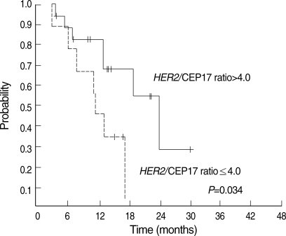

그림 출처: PubMed Central 공개 논문 (PMC2752777)
그 밖의 정보
- 출판 형태
- Journal Article, Research Support, Non-U.S. Gov't
- 저널
- Journal of Korean medical science · eISSN 1598-6357
- 전자 출판
- 2009-09-24
- MeSH
- Adult · Aged · Antibodies, Monoclonal / administration & dosage / therapeutic use · Antibodies, Monoclonal, Humanized · Antineoplastic Combined Chemotherapy Protocols / administration & dosage / therapeutic use · Breast Neoplasms / drug therapy / mortality / pathology · Disease Progression · Drug Administration Schedule · Female · Gene Amplification · Humans · In Situ Hybridization, Fluorescence · Middle Aged · Paclitaxel / administration & dosage / therapeutic use · Predictive Value of Tests · Erb-b2 Receptor Tyrosine Kinases / genetics / metabolism · Survival Analysis · Trastuzumab
- Substances
- Antibodies, Monoclonal · Antibodies, Monoclonal, Humanized · Paclitaxel · Erb-b2 Receptor Tyrosine Kinases · Trastuzumab
- 참고문헌
- 25편