SCI 발표논문

SCI 발표논문

← SCI 발표논문 목록

Respir Res. 2023;24(1):68.

Classical monocyte-derived macrophages as therapeutic targets of umbilical cord mesenchymal stem cells: comparison of intratracheal and intravenous administration in a mouse model of pulmonary fibrosis.

, , , , , , , ,

제1저자 최선미, 모요셉 · 교신저자 강혜련

소속 (10)
  1. Division of Pulmonary and Critical Care Medicine, Department of Internal Medicine, Seoul National University Hospital, Seoul, Republic of Korea.
  2. Department of Translational Medicine, Seoul National University College of Medicine, Seoul, Republic of Korea.
  3. Institute of Allergy and Clinical Immunology, Seoul National University Medical Research Center, Seoul, Republic of Korea.
  4. Laboratory of Mucosal Immunology, Department of Biomedical Sciences, Seoul National University College of Medicine, Seoul, Republic of Korea.
  5. Department of Critical Care Medicine, Seoul National University Hospital, Seoul, Republic of Korea.
  6. Department of Pathology, Seoul National University College of Medicine, Seoul, Republic of Korea.
  7. Department of Internal Medicine, Seoul National University College of Medicine, Seoul, Republic of Korea.
  8. Department of Translational Medicine, Seoul National University College of Medicine, Seoul, Republic of Korea. helenmed@snu.ac.kr.
  9. Department of Internal Medicine, Seoul National University College of Medicine, Seoul, Republic of Korea. helenmed@snu.ac.kr.
  10. Institute of Allergy and Clinical Immunology, Seoul National University Medical Research Center, Seoul, Republic of Korea. helenmed@snu.ac.kr.

Abstract

BACKGROUND Idiopathic pulmonary fibrosis (IPF) is a progressive fibrotic lung disease that has no cure. Although mesenchymal stem cells (MSCs) have been reported to ameliorate lung inflammation and fibrosis in mouse models, their mechanisms of action remain unknown. Therefore, we aimed to determine the changes in various immune cells, especially macrophages and monocytes, involved in the effects of MSC treatment on pulmonary fibrosis.

METHODS We collected and analyzed explanted lung tissues and blood from patients with IPF who underwent lung transplantation. After establishing a pulmonary fibrosis model via the intratracheal administration of bleomycin (BLM) to 8-week-old mice, MSCs derived from human umbilical cords were administered intravenously or intratracheally on day 10 and the lungs were immunologically analyzed on days 14 and 21. Flow cytometry was performed to analyze the immune cell characteristics, and gene expression levels were examined using quantitative reverse transcription-polymerase chain reaction.

RESULTS In the histological analysis of explanted human lung tissues, the terminally fibrotic areas contained a larger number of macrophages and monocytes than the early fibrotic areas of the lungs. When human monocyte-derived macrophages (MoMs) were stimulated with interleukin-13 in vitro, the expression of type 2 macrophage (M2) markers was more prominent in MoMs from the classical monocyte subset than in those from intermediate or non-classical monocyte subsets, and MSCs suppressed M2 marker expression independent of MoM subsets. In the mouse model, the increased number of inflammatory cells in the bronchoalveolar lavage fluid and the degree of lung fibrosis observed in BLM-treated mice were significantly reduced by MSC treatment, which tended to be more prominent with intravenous administration than intratracheal administration. Both M1 and M2 MoMs were upregulated in BLM-treated mice. The M2c subset of M2 MoMs was significantly reduced by MSC treatment. Among M2 MoMs, M2 MoMs derived from Ly6C+ monocytes were most effectively regulated by the intravenous administration, not intratracheal administration, of MSCs.

CONCLUSIONS Inflammatory classical monocytes may play a role in lung fibrosis in human IPF and BLM-induced pulmonary fibrosis. Intravenous rather than intratracheal administration of MSCs may ameliorate pulmonary fibrosis by inhibiting monocyte differentiation into M2 macrophages.

© 2023. The Author(s).

Keywords

BleomycinMacrophageMesenchymal stem cellMonocytePulmonary fibrosis

Figures
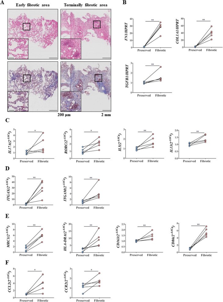
Fig. 1
Fig. 1 Pathophysiologic changes in samples of patients with IPF. A H&E stain and MT stain (× 40 and × 100) of lung tissue of patients with IPF. B Changes in FN1, COL1A1, and TGB1 mRNA expression in lungs of patients with IPF. C Changes in IL17A, RORC, IL5and IL13 mRNA expression in lungs of patients with IPF. D Changes in ITGAX and ITGAM mRNA expression in lungs of patients with IPF. E MRC1, HLA-DRA CD163, and CD86 mRNA expression in lungs of patients with IPF. F CCL2 and CCR2 mRNA expression in lungs of patients with IPF. n = 5 for each group, *indicates P < 0.05, **indicates P < 0.01. All results are representative of at least three independent experiments. IPF idiopathic pulmonary fibrosis, H&E hematoxylin and eosin, MT Masson's trichrome, IL interleukin
Fig. 2
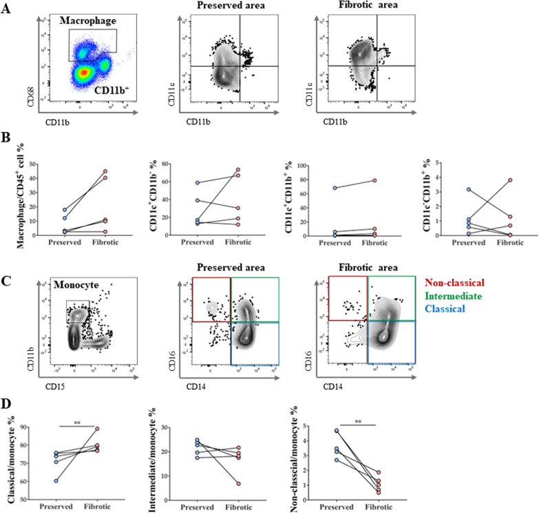
Fig. 2 Monocyte and macrophage subtype changes in IPF patient lungs. A Macrophage gating protocol and dot plots showing lung AM and MoM distribution in vivo according to CD11c versus CD11b marker expression. B Changes in total macrophages and their subtypes in lungs of patients with IPF. C Gating protocol of monocytes and their subtypes. D Changes in monocyte subtype in lungs of patients with IPF. n = 5 for each group, *indicates P < 0.05, **indicates P < 0.01. All results are representative of at least three independent experiments. IPF idiopathic pulmonary fibrosis, AM alveolar macrophages, MoM monocyte-derived macrophage
Fig. 3
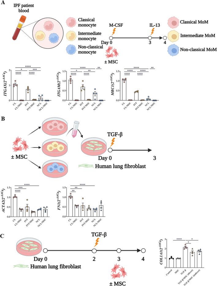
Fig. 3 Confirmation of lung fibrosis induction by subtypes of monocyte and macrophage. A Monocyte subtype isolation and macrophage differentiation protocol and their macrophage differentiation ability and M2 activation ability B Monocyte-derived macrophage culture media transfer protocol and the ability of monocyte subtypes to induce fibrosis. *indicates P < 0.05, **indicates P < 0.01, ***indicates P < 0.001, ****indicates P<0.0001
Fig. 4
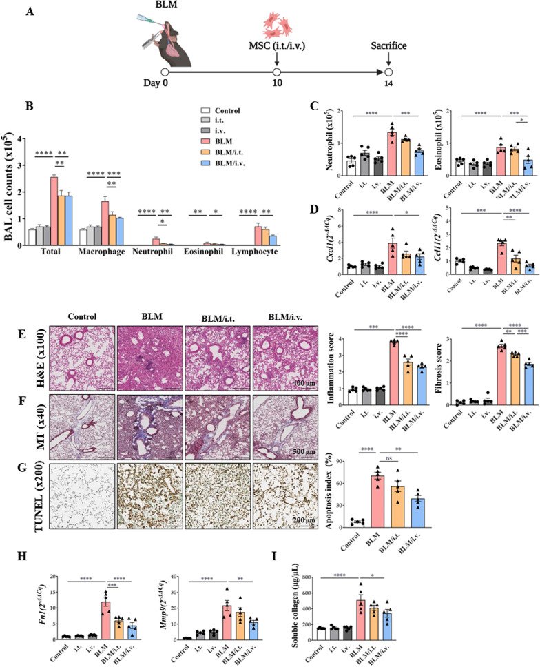
Fig. 4 Therapeutic effect of MSCs in a murine fibrosis model on the modulation of exacerbated inflammation. A The murine BLM-induced fibrosis model used in the study; administration of intratracheal BLM at 3 mg/kg on day 0. Intratracheal or intravenous treatment of MSCs (105 cells) on day 10. B The change of immune cells including macrophages, neutrophils, eosinophils, and lymphocytes in BAL fluid. C The number of neutrophils and eosinophils in lungs analyzed by flow cytometry. D Chemotaxis mRNA changes associated with neutrophils and eosinophils in the lung. E H&E stain (× 100) of lung histology and their scoring; Inflammation score: 0 (absent), 1 (discrete), 2 (mild), 3 (moderate), and 4 (intense). F MT stain (× 40) of lung histology and their scoring; Fibrosis score: 0 (none), 1 (mild), 2 (moderate), and 3 (severe). G TUNEL stain (× 200) of lung histology and their scoring; Apoptosis index (%): Quantification of the TUNEL-staining cells. H Fn1 and Mmp9 expression in lungs. I Result of Sircol assay showing changes in soluble collagen concentration in the lungs. n = 5 for each group, *indicates P < 0.05, **indicates P < 0.01, ***indicates P < 0.001, ****indicates P < 0.0001. All results are representative of at least three independent experiments. MSC mesenchymal stem cell, BLM bleomycin, BAL bronchoalveolar lavage, H&E hematoxylin and eosin, MT Masson's trichrome, TUNEL terminal deoxynucleotidyl transferase dUTP nick-end labeling, Fn fibronectin, Mmp9 matrix metalloproteinase-9
Fig. 5
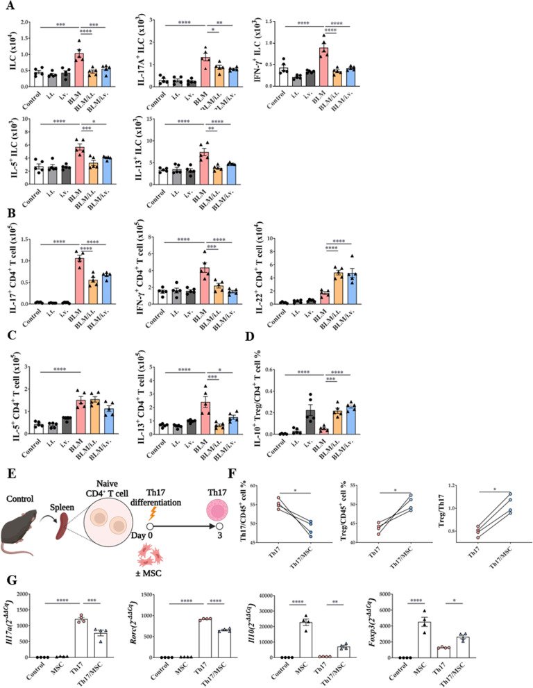
Fig. 5 Modulating effect of MSCs on ILCs and T cells activation in a BLM-induced lung fibrosis model. A The number of ILCs, IL-17A+ ILCs, IFN-γ+ ILCs, IL-5+ ILCs and IL-13+ ILCs in lungs. B The number of IL-17A+ CD4+ T cells, IFN-γ+ CD4+ T cells, and IL-22+ CD4+ T cells in lungs. C The number of IL-5+ CD4+ T cells and IL-13+ CD4+ T cells in lungs. D The ratio of IL-10+ Tregs in lungs. E Th17 differentiation protocol. F The ratio of Th17 and Treg. G Changes in Il17, Rorc, Il10, and Foxp3 mRNA expression by MSCs in the Th17 cells. n = 4–5 for each group, *indicates P < 0.05, **indicates P < 0.01, ***indicates P < 0.001, ****indicates P < 0.0001. All results are representative of at least three independent experiments. ILC innate lymphoid cell, IL interleukin, IFN interferon, Th17 T helper 17 cells; Treg, regulatory T cells
Fig. 6
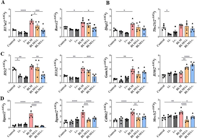
Fig. 6 Effect of MSCs on mRNA expression of lung in a murine fibrosis model. A–C Changes in Il17a, Rorc,Ifng, Tbx21, Il5, Il13, Gata3, and Il10 mRNA expression by MSCs in the lungs. D Changes in Itgax, Igtam, Mrc1, and Cd86 mRNA expression by MSCs in the lungs. All data were analyzed via real-time quantitative PCR analysis. n = 5 for each group, *indicates P < 0.05, **indicates P < 0.01, ***indicates P < 0.001, ****indicates P < 0.0001. All results are representative of at least three independent experiments. MSC mesenchymal stem cell, IL interleukin, IFN interferon
Fig. 7
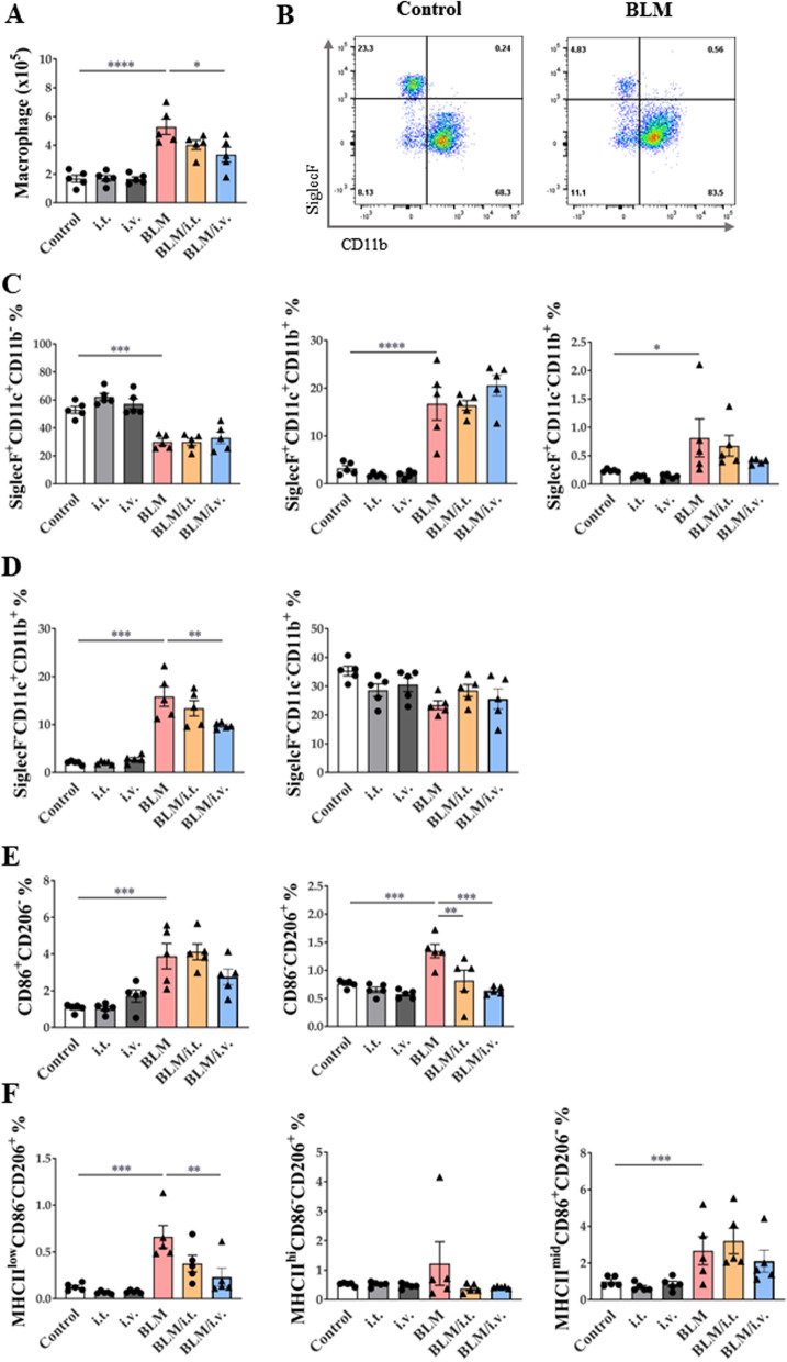
Fig. 7 Changes in macrophage subtype activation by MSCs in a murine fibrosis model. A The number of lung macrophages. B Dot plots showing lung AM and MoM distribution in vivo according to SiglecF versus CD11b marker expression. C, D Subtypes of SiglecF+ and SiglecF– lung macrophages according to CD11c and CD11b expression. E Changes in MoMs in the lungs and expression of CD86 and CD206 markers in MoMs (F) Changes in M2 macrophage subpopulations according to the expression of MHCII, CD206, and CD86 markers in the lungs. n = 5 for each group, *indicates P < 0.05, **indicates P < 0.01, ***indicates P < 0.001, ****indicates P < 0.0001. All results are representative of at least three independent experiments. MSC mesenchymal stem cell, AM alveolar macrophages, MoM monocyte-derived macrophage
Fig. 8
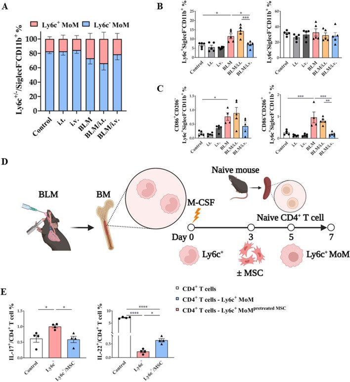
Fig. 8 Regulatory effect of MSCs on in vivo and ex vivo Ly6c+/– macrophages activation. A, B Change of Ly6c+ and Ly6c− MoMs in lungs. C Changes in CD86 and CD206 marker expression populations in Ly6c+ MoMs in the lungs. D Ex vivo model to evaluate the regulation effect of Ly6c+ MoM on CD4+ T cell plasticity and the effect of MSC treatment on the regulation E Effect of MSC on the plasticity of naive CD4+ T cells co-cultured with Ly6c+ MoMs. n = 4–5 for each group, *indicates P < 0.05, **indicates P < 0.01, ***indicates P < 0.001, ****indicates P < 0.0001. All results are representative of at least three independent experiments. MSC mesenchymal stem cell, MoM monocyte-derived macrophage
Fig. 9
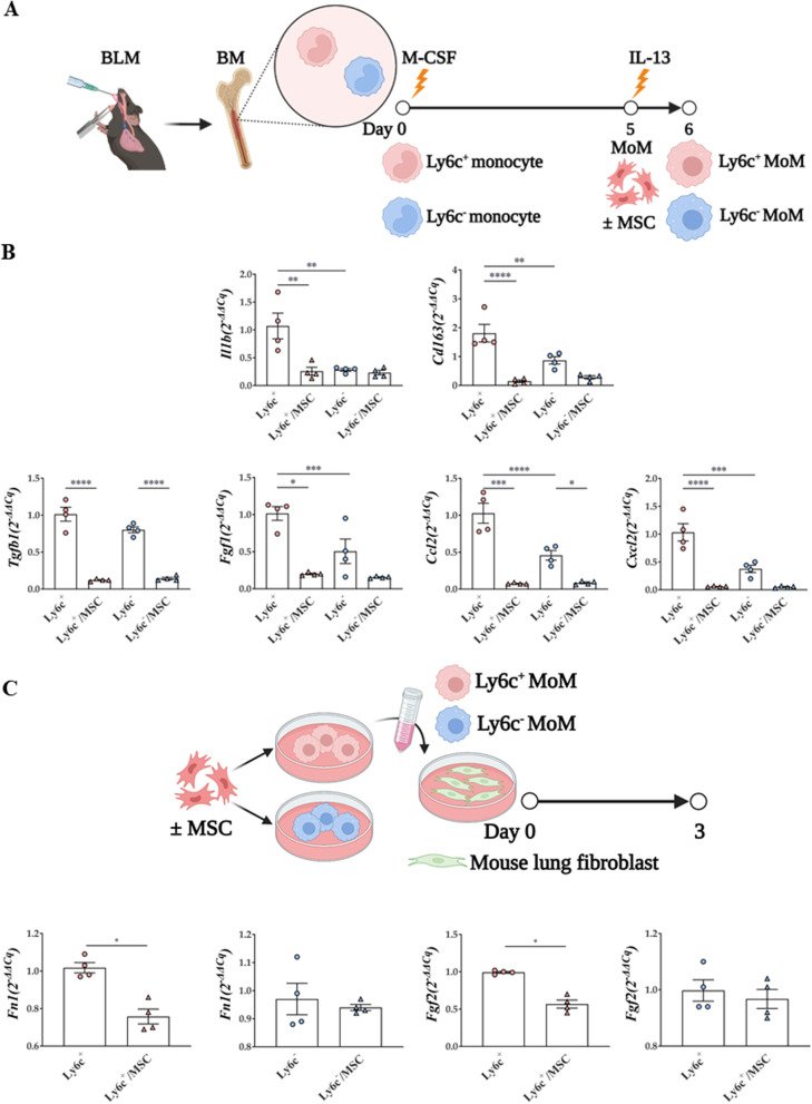
Fig. 9 Regulatory effect of MSCs on and ex vivo and in vitro Ly6c+/– macrophages activation. A Macrophage differentiation protocol of Ly6c+ or Ly6c– monocytes isolated from the BLM model. B mRNA levels of macrophage activation markers, fibrosis-related markers, and immune cell chemotaxis markers in Ly6c+ or Ly6c–MoMs. C Comparison of fibrosis-inducing ability of Ly6c+ or Ly6c– MoMs. n = 4–5 for each group, *indicates P < 0.05, **indicates P < 0.01, ***indicates P < 0.001, ****indicates P < 0.0001. All results are representative of at least three independent experiments. MSC mesenchymal stem cell, BLM bleomycin, MoM monocyte-derived macrophage

그림 출처: PubMed Central 공개 논문 (PMC9985859)

Conflict of interest statement

The authors declare that they have no competing interests.

그 밖의 정보
출판 형태
Journal Article
저널
Respiratory research · eISSN 1465-993X
전자 출판
2023-03-05
MeSH
Humans · Animals · Mice · Administration, Intravenous · Macrophages · Monocytes · Idiopathic Pulmonary Fibrosis* · Bleomycin · Disease Models, Animal · Mesenchymal Stem Cells*
Substances
Bleomycin
참고문헌
89편