
J Cardiovasc Magn Reson. 2020;22(1):25.
Myocardial structural and functional changes in patients with liver cirrhosis awaiting liver transplantation: a comprehensive cardiovascular magnetic resonance and echocardiographic study.
제1저자 김혜미 · 교신저자 김형관, 이정훈
소속 (6)
- Division of Cardiology, Department of Internal Medicine and Cardiovascular Center, Section of Cardiovascular Imaging, Seoul National University Hospital, 103 Daehak-ro, Jongno-gu, Seoul, 03080, South Korea.
- Division of Cardiology, Department of Internal Medicine, Chung-Ang University Hospital, Seoul, South Korea.
- Division of Cardiology, Department of Internal Medicine and Cardiovascular Center, Section of Cardiovascular Imaging, Seoul National University Hospital, 103 Daehak-ro, Jongno-gu, Seoul, 03080, South Korea. cardiman73@gmail.com.
- Division of Gastroenterology, Department of Internal Medicine and Liver Research Institute, Seoul National University College of Medicine, 103 Daehak-ro, Jongno-gu, Seoul, 03080, South Korea. pindra@empal.com.
- Division of Gastroenterology, Department of Internal Medicine and Liver Research Institute, Seoul National University College of Medicine, 103 Daehak-ro, Jongno-gu, Seoul, 03080, South Korea.
- Department of Radiology, Seoul National University Hospital, Seoul, South Korea.
Abstract
BACKGROUND Cardiac dysfunction is increasingly recognized in patients with liver cirrhosis. Nevertheless, the presence or absence of structural alterations such as diffuse myocardial fibrosis remains unclear. We aimed to investigate myocardial structural changes in cirrhosis, and explore left ventricular (LV) structural and functional changes induced by liver transplantation.
METHODS This study included 33 cirrhosis patients listed for transplantation and 20 healthy controls. Patients underwent speckle-tracking echocardiography and cardiovascular magnetic resonance (CMR) with extracellular volume fraction (ECV) quantification at baseline (n = 33) and 1 year after transplantation (n = 19).
RESULTS CMR-based LV ejection fraction (CMRLV-EF) and echocardiographic LV global longitudinal strain (LV-GLS) demonstrated hyper-contractile LV in cirrhosis patients (CMRLV-EF: 67.8 ± 6.9% in cirrhosis vs 63.4 ± 6.4% in healthy controls, P = 0.027; echocardiographic GLS: - 24.2 ± 2.7% in cirrhosis vs - 18.6 ± 2.2% in healthy controls, P < 0.001). No significant differences in LV size, wall thickness, mass index, and diastolic function between cirrhosis patients and healthy controls were seen (all P > 0.1). Only one of the cirrhosis patients showed late gadolinium enhancement. However, cirrhosis patients showed a higher ECV (31.6 ± 5.1% vs 25.4 ± 1.9%, P < 0.001) than healthy controls. ECV showed a positive correlation with Child-Pugh score (r = 0.564, P = 0.001). Electrocardiogram-based corrected QT interval was prolonged in cirrhosis (P < 0.001). One-year post-transplantation, echocardiographic LV-GLS (from - 24.9 ± 2.4% to - 20.6 ± 3.4%, P < 0.001) and ECV (from 30.9 ± 4.5% to 25.4 ± 2.6%, P = 0.001) moved to the normal ranges. Corrected QT interval decreased after transplantation (from 475 ± 41 to 429 ± 30 msec, P = 0.001).
CONCLUSIONS Myocardial extracellular volume expansion with augmented resting LV systolic function was characteristic of cirrhotic cardiomyopathy, which normalizes 1-year post-transplantation. Thus, myocardial extracellular expansion represents a structural component of myocardial changes in cirrhosis.
Keywords
Cardiovascular magnetic resonanceCirrhosisExtracellular volume fractionGlobal longitudinal strainLeft ventricle
Figures
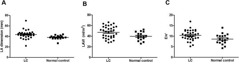
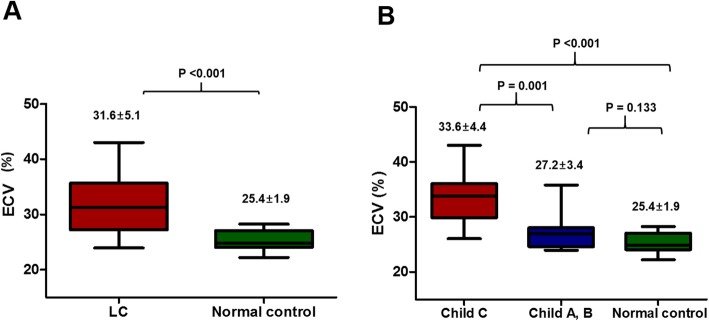
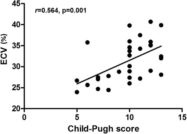
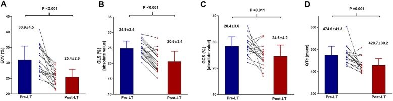
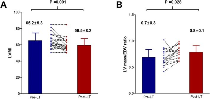
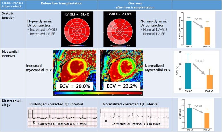
그림 출처: PubMed Central 공개 논문 (PMC7178724)
Conflict of interest statement
Dr. Hyung-Kwan Kim reports research grants from Actelion, Handok Pharmaceuticals, Dae-Woong Pharmaceuticals, and Norvatis. Dr. Jung-Hwan Yoon reports research grants from Bayer HealthCare Pharmaceuticals and AstraZeneca; and Dr. Yoon Jun Kim, research grants from BTG, Bayer HealthCare Pharmaceuticals, Ono, AstraZeneca, Roche, LG Life Science, and Bristol-Myers Squibb, lecture fees from Bayer HealthCare Pharmaceuticals and Gilead Science, and serving as an advisory board member or consultant of Gilead Science, Bayer HealthCare Pharmaceuticals, Ono, and AbbVie. All other authors report no conflict of interest.
그 밖의 정보
- 출판 형태
- Journal Article, Research Support, Non-U.S. Gov't
- 저널
- Journal of cardiovascular magnetic resonance : official journal of the Society for Cardiovascular Magnetic Resonance · eISSN 1532-429X
- 전자 출판
- 2020-04-23
- MeSH
- Aged · Cardiomyopathies / diagnostic imaging / etiology / physiopathology · Case-Control Studies · Echocardiography, Doppler* · Female · Humans · Hypertrophy, Left Ventricular / diagnostic imaging / etiology / physiopathology · Liver Cirrhosis / complications / diagnostic imaging / surgery · Liver Transplantation* · Magnetic Resonance Imaging, Cine* · Male · Middle Aged · Predictive Value of Tests · Prospective Studies · Recovery of Function · Time Factors · Treatment Outcome · Ventricular Dysfunction, Left / diagnostic imaging / etiology / physiopathology · Ventricular Function, Left · Ventricular Remodeling · Waiting Lists*
- 참고문헌
- 44편