Immunoregulation Section, Autoimmunity Branch, NIAMS, NIH, Bethesda, MD, USA.
Division of Rheumatology, Department of Internal Medicine, MMBS, Medical Research Center, Seoul National University College of Medicine, Seoul, Korea.
Division of Rheumatology, University of Colorado School of Medicine, Aurora, CO, 80207, USA.
Division of Rheumatology, Department of Medicine, Los Angeles County University of Southern California Medical Center and University of Southern California Keck School of Medicine, Los Angeles, CA, USA.
Rheumatology Division, Department of Medicine, University of Nebraska Medical Center and VA Nebraska-Western Iowa Health Care System, Omaha, NE, USA.
Liver Diseases Branch, NIDDK, NIH, Bethesda, MD, USA.
Human Genome Sciences, Rockville, MD, USA.
Immunology Biomarkers group, Pharmaceutical Companies of J&J, LLC, Spring House, PA, USA.
Clinical Trials and Outcomes Branch, NIAMS, NIH, Bethesda, MD, USA.
Brigham and Women's Hospital, Harvard Medical School, Boston, MA, USA.
Present address: Janssen Research and Development, Spring House, PA, USA.
Colorado School of Public Health, Aurora, CO, USA.
Center for Genomics & Human Genetics, The Feinstein Institute for Medical Research, Hofstra North Shore-LIJ School of Medicine, Manhasset, NY, USA.
Immunoregulation Section, Autoimmunity Branch, NIAMS, NIH, Bethesda, MD, USA. richardmsiegel@gmail.com.
Abstract
BACKGROUND The tumor necrosis factor (TNF) superfamily cytokine TNF-like protein 1A (TL1A) and its receptor DR3 are essential for diverse animal models of autoimmune disease and may be pathogenic in rheumatoid arthritis (RA). However, the relationship of TL1A to disease duration, activity, and response to anti-TNF and other therapies in RA is not clear.
METHODS We measured soluble TL1A in synovial fluid (SF), serum, or plasma from RA first-degree relatives (FDRs) and in early RA and established disease. We measured the effects of anti-TNF and methotrexate (MTX) therapy on circulating TL1A from multiple independent RA treatment trials. We also determined the ability of a blocking anti-TL1A antibody to inhibit clinical disease and articular bone destruction in the murine collagen-induced arthritis (CIA) model of human RA.
RESULTS Soluble TL1A was specifically elevated in the blood and SF of patients with RA compared to patients with other diseases and was elevated early in disease and in at-risk anti-cyclic citrullinated peptide (CCP) (+) first-degree relatives (FDRs). Therapeutic TNF inhibition reduced serum TL1A in both responders and non-responders, whereas TL1A declined following MTX treatment only in responders. In murine CIA, TL1A blockade was clinically efficacious and reduced bone erosions.
CONCLUSIONS TL1A is specifically elevated in RA from early in the disease course and in at-risk FDRs. The decline in TL1A after TNF blockade suggests that TL1A levels may be a useful biomarker for TNF activity in RA. These results support the further investigation of the relationship between TL1A and TNF and TL1A blockade as a potential therapeutic strategy in RA.
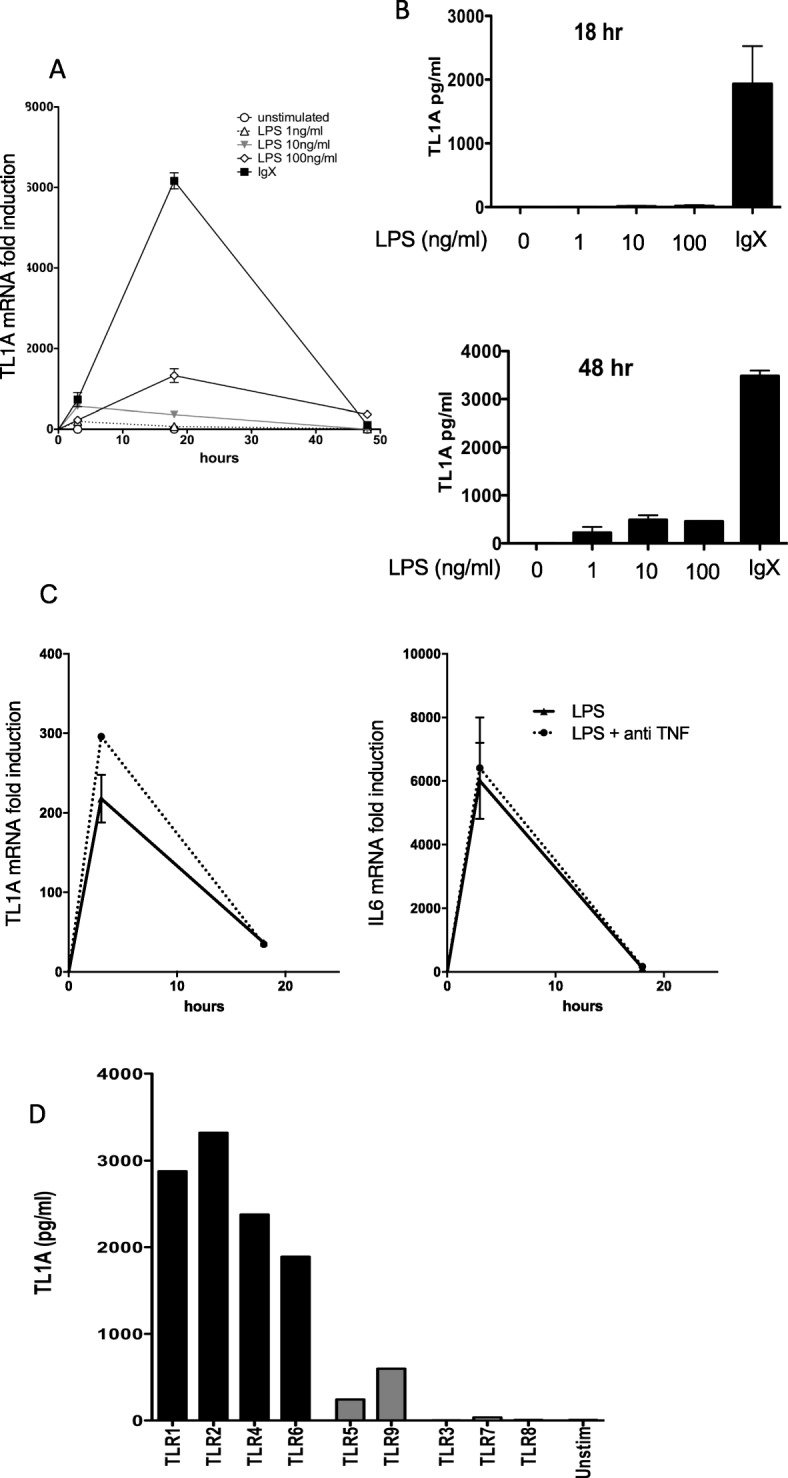
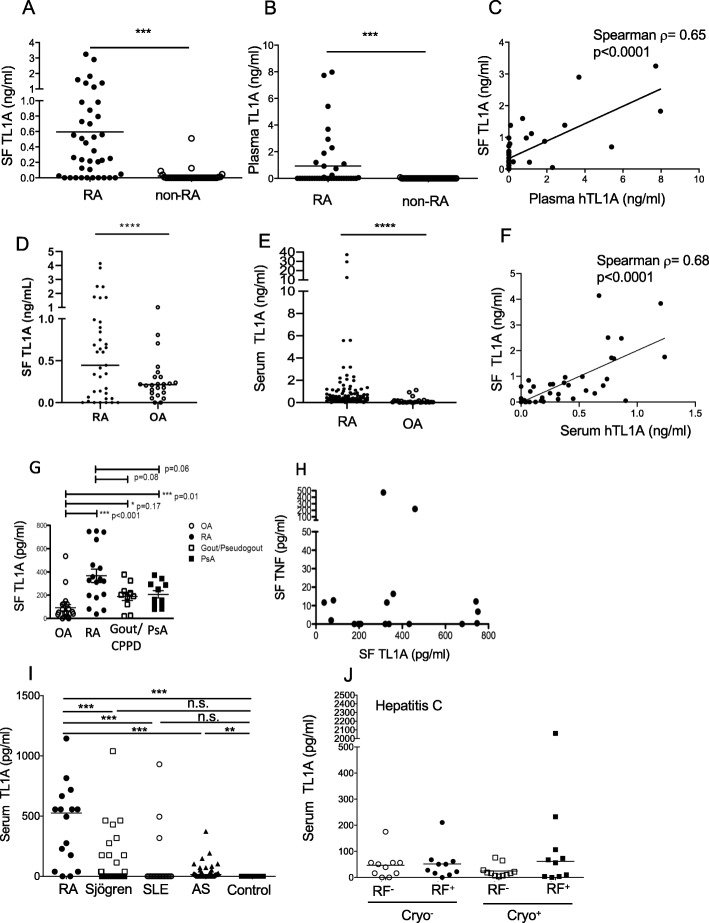
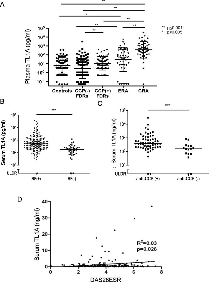
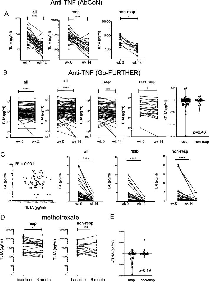
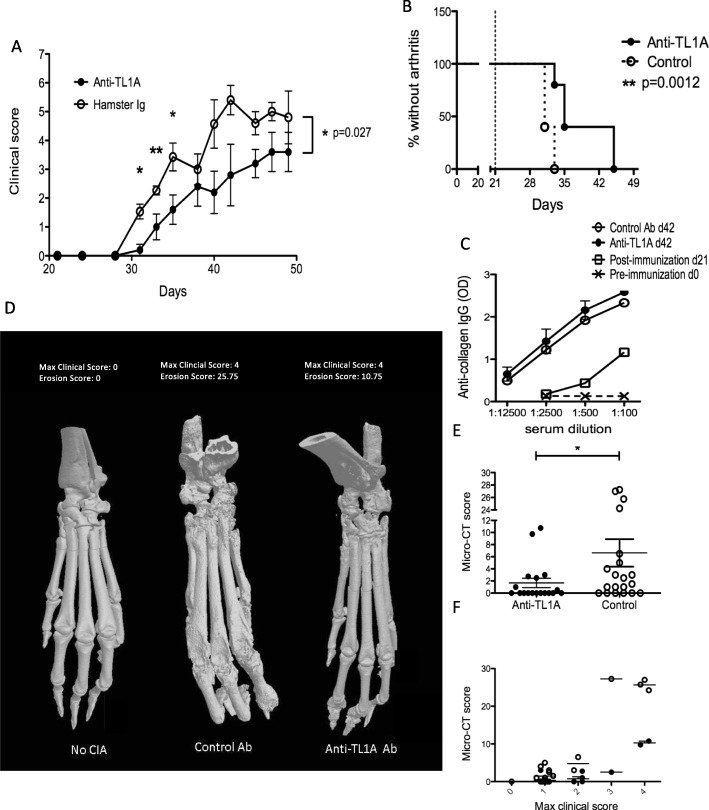
Fig. 1Induction of TL1A by immune stimuli in human monocytes. a Human peripheral blood monocytes (1 × 10⁶ cells/ml) were cultured with the indicated concentrations of LPS or with plate-bound immune complexes (IgX). Total RNA was isolated at the indicated time points, and TL1A gene expression was measured using qRT-PCR. TL1A mRNA levels were measured relative to human β2-microglobulin and normalized to that in resting monocytes and are shown as mean ± s.e.m. Data is representative of 5 independent experiments with different donors. b Monocyte culture supernatants were collected at 18 h and 48 h, and TL1A levels were measured using ELISA as described in the methods. Results in each panel are mean ± s.e.m. and are representative of a minimum of three experiments. Data is representative of results from 5 (LPS) and 3 (IgX) independent donors. c Peripheral blood monocytes were treated as in a with (100 ng/ml) LPS in the presence or absence of anti-TNF (10 μg/ml) and induction of TL1A and IL-6 measured by qRT-PCR. Data is representative of two independent experiments with different donors. d Monocytes at a higher initial concentration (2 × 10⁶ cells/ml) were stimulated with the indicated TLR agonist, and TL1A was measured in the supernatant after 48 h with a bead-based assay as described in the methods. Results are from one of two independent experiments with different donorsFig. 2TL1A elevation in rheumatoid arthritis and other diseases. a Soluble TL1A was measured in SF from patients diagnosed with RA (n = 39) or other types of arthritis (non-RA, n = 37) including psoriatic arthritis, crystal-induced arthritis, reactive arthritis, non-inflammatory arthritis, nonspecific inflammatory arthritis, JIA, or infectious arthritis. Values are shown with a line representing the median value and significance of the difference between RA and non-RA samples p ≤ 0.0001 (Mann-Whitney test). b TL1A was measured in matching blood plasma samples as in a. p = 0.0002 (Mann-Whitney test). c TL1A levels in matched synovial fluid and plasma samples from RA patients (n = 39) were compared. Non-parametric correlation of SF vs. plasma TL1A is shown with the indicated Spearman’s ρ and p value. d Soluble TL1A was measured in synovial fluid (SF) from patients diagnosed with RA (n = 34) and osteoarthritis (OA) (n = 27). Values are shown with a line representing the median value and significance of the difference between RA and OA samples. p = 0.143 (Mann-Whitney test). e TL1A was measured in serum samples from patients diagnosed with RA (n = 98) and OA (n = 30) ****p ≤ 0.0001 (Mann-Whitney test). f TL1A levels in matched synovial fluid and serum samples from RA patients (n = 34) were compared. One outlier with very high TL1A in both serum (5.6 ng/ml) and SF (8.4 ng/ml) was removed from the analysis. Non-parametric correlation of SF vs. serum TL1A is shown with the indicated Spearman’s ρ and p value. For all panels. g TL1A was measured in SF from patients diagnosed with the indicated diseases using a bead-based immunoassay as described in the methods. RA n = 17; OA, n = 21; gout/CPPD (calcium pyrophosphate disease), n = 10; PsA (psoriatic arthritis), n = 11. Asterisks represent significances compared to OA (Mann-Whitney test) *p ≤ 0.05, ***p ≤ 0.001. h TL1A and TNF levels in SF from RA patients were measured by a bead-based assay as described in the methods. i Serum TL1A was measured as in g from patients diagnosed with the indicated diseases: RA, n = 20; AS ankylosing spondylitis, n = 40; Sjögren’s syndrome, n = 59; SLE systemic lupus erythematosus, n = 20; control, n = 10. Asterisks represent p value compared to control group **p ≤ 0.01, ***p ≤ 0.001 (Mann-Whitney test). j TL1A was measured in the serum of patients with hepatitis C with the indicated status for circulating cryoglobulins and/or rheumatoid factorFig. 3Relationship between circulating TL1A and disease duration, serological markers, and clinical activity in RA. a Plasma TL1A measured in the indicated groups of subjects from the SERA cohort. FDR: first-degree relatives grouped into those with or without anti-CCP antibodies. ERA, early RA (< 1 year); CRA, chronic RA (> 1 year). Significance of differences given by Mann-Whitney test with Bonferroni correction for multiple testing. Serum TL1A in RA patients from the SNUH cohort grouped by RF positivity (b) and anti-CCP positivity (c). Significance of the difference given by Mann-Whitney test. *p ≤ 0.05, **p ≤ 0.01,***p ≤ 0.001, ****p ≤ 0.0001. d Linear correlation of serum TL1A and DAS28 CRP in the SNUH RA cohortFig. 4Effects of anti-TNF and MTX treatment on TL1A levels in rheumatoid arthritis. a Soluble TL1A was measured in serum samples from the AbCoN cohort [20] before or 14 weeks after treatment with TNF inhibitors. Subjects were classified by EULAR criteria into responders (good and moderate response, n = 34) and non-responders, n = 15. Significance for a change in TL1A was measured by Wilcoxon signed rank analysis. b Similar analysis was performed in a larger cohort (n = 100) of RA patients treated with golimumab, with serum TL1A levels shown at baseline compared to week 2 in all patients and week 14 in all patients, and grouped into responders (76) and non-responders (24) by EULAR criteria as in a. The right panel shows change in TL1A (14 weeks baseline) compared between responders and non-responders. The significance of the difference in ΔTL1A between responders and non-responders is given (Mann-Whitney non-parametric test). c Serum IL-6 measured in the same cohort of RA patients. Correlation with TL1A levels at baseline (left) and IL-6 levels at baseline and week 14 in all patients, and grouped into responders and non-responders by EULAR criteria as in b (right). d TL1A was measured in the serum before (baseline) or after 6 months of treatment with MTX monotherapy (17.5 mg/week) in 41 patients with early RA. Responsiveness was measured by EULAR criteria as described in the methods (23 good/moderate responders, 18 non-responders). e The change in TL1A (6 months baseline) was compared between responders and non-responders identified in d. The significance of the difference in ΔTL1A between responders and non-responders is given (Mann-Whitney non-parametric test)Fig. 5Blocking TL1A-DR3 interactions prevents disease and bone erosions in collagen-induced arthritis. a CIA was induced in DBA/1 mice as described in the methods. Weekly intra-peritoneal injections of 20 mg/kg of either anti-TL1A (treatment, n = 5) or hamster immunoglobulin Ig control (n = 7) were begun at day 21 after initial immunization with collagen. A representative experiment of three independent experiments is shown. Clinical scores on each day were compared using an unpaired t test, and p values for significance are shown above each time-point represented by asterisks (*p < 0.05, **p < 0.01) above each day. Two-way ANOVA was also performed to compare the trend of the two graphs, with p value shown to the right. b Survival analysis of the percentage of mice without arthritis on each day is compared between the anti-TL1A treated group and control group. Arthritis was defined by a combined clinical score of two or more. c Sera from mice from each group induced to develop CIA as in a were collected at indicated time points and anti-chicken collagen IgG levels were measured by ELISA. d 3-D reconstructions of micro-CT examination of hind paws from mice induced to develop CIA with and without TL1A blockade. Examples are shown from each treatment group, with the maximal clinical scores and the erosion score obtained for that paw by two separate observers blinded to treatment groups. e Composite of CT erosion scores obtained from the anti-TL1A treated group (n = 18) and control-antibody treated group (n = 20), significance is from an unpaired t test with Welch’s correlation (*p ≤ 0.05). Analysis of individual regions resulted in p values of 0.078 at ankle/tarsus, 0.042 at metatarsophalangeal (MTP) joints, and 0.015 at toes. f Comparison of the CT scores of the paws from the two groups based on the maximum clinical scores. Anti-TL1A treatment significantly reduced erosions independent of the clinical score. p < 0.0001 by 2-way ANOVA
그림 출처: PubMed Central 공개 논문 (PMC7204024)
Conflict of interest statement
Chris D. Ward and Thi-Sau Migone were employees of Human Genome Sciences and owners of HGSI stock. Sarah Lamberth and David Lee are employees of Janssen Pharmaceuticals and own stock. V. Michael Holers and Kevin D. Deane are co-holders of a Stanford University patent on use of biomarkers to predict RA development. Richard Siegel, Yun-Jeong Song, and Françoise Meylan hold a patent on anti-TL1A antibodies. Richard Siegel has been an employee of Novartis Institutes of Biomedical Research since 2018 and owns Novartis stock. The remaining authors declared no conflict of interest.
그 밖의 정보
출판 형태
Journal Article, Research Support, N.I.H., Extramural, Research Support, N.I.H., Intramural, Research Support, Non-U.S. Gov't