Strategic Center of Cell and Bio Therapy for Heart, Diabetes & Cancer, Biomedical Research Institute, Seoul National University Hospital, Seoul 03080, Republic of Korea.
Stem Cell Convergence Research Center, Korea Research Institute of Bioscience and Biotechnology (KRIBB), 125 Gwahak-ro, Yuseong-gu, Daejeon 34141, Republic of Korea.
Cardiovascular Center & Department of Internal Medicine, Seoul National University Hospital, Seoul 03080, Republic of Korea.
ToolGen, Inc., #1204, Byucksan Digital Valley 6-cha, 219 Gasan Digital 1-ro, Geumcheon-gu, Seoul 08501, Republic of Korea.
SK Chemical, Life Science Business, Clinical Research Team, Gyeonggi-do, 13494, Republic of Korea.
GenNBio, Inc., 422, Teheran-ro, Gangnam-gu, Seoul 06193, Republic of Korea.
Molecular Medicine and Biopharmaceutical Sciences, Graduate School of Convergence Science and Technology, Seoul National University, Seoul 03080, Republic of Korea.
Abstract
Although human induced pluripotent stem cells (iPSCs) can serve as a universal cell source for regenerative medicine, the use of iPSCs in clinical applications is limited by prohibitive costs and prolonged generation time. Moreover, allogeneic iPSC transplantation requires preclusion of mismatches between the donor and recipient human leukocyte antigen (HLA). We, therefore, generated universally compatible immune nonresponsive human iPSCs by gene editing. Transcription activator-like effector nucleases (TALENs) were designed for selective elimination of HLA DR expression. The engineered nucleases completely disrupted the expression of HLA DR on human dermal fibroblast cells (HDF) that did not express HLA DR even after stimulation with IFN-γ. Teratomas formed by HLA DR knockout iPSCs did not express HLA DR, and dendritic cells differentiated from HLA DR knockout iPSCs reduced CD4+ T cell activation. These engineered iPSCs might provide a novel translational approach to treat multiple recipients from a limited number of cell donors.
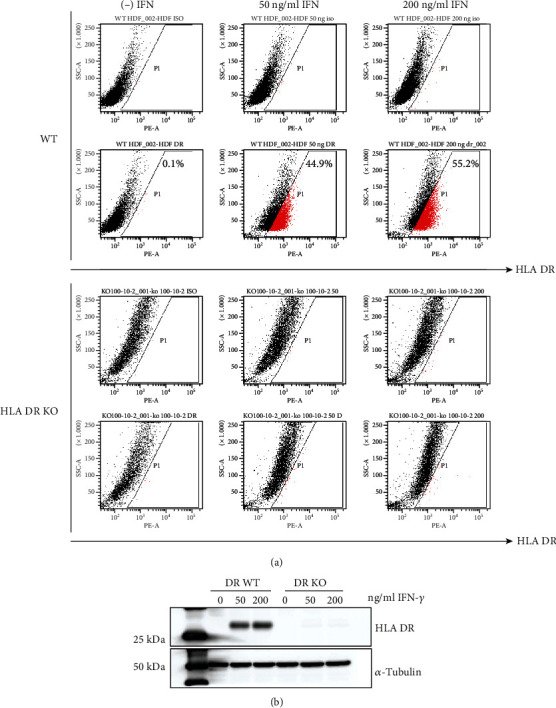
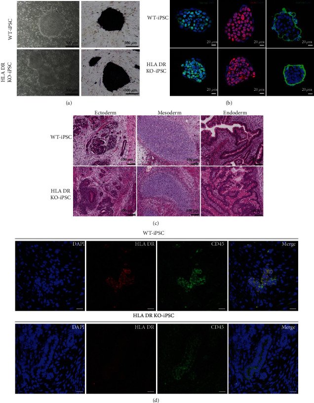
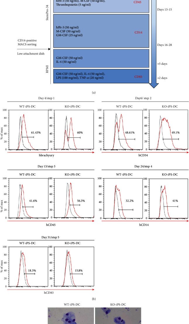
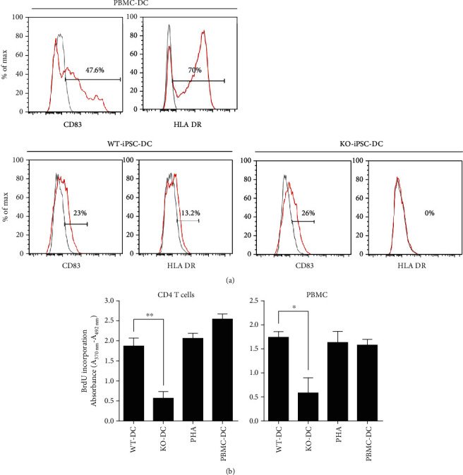
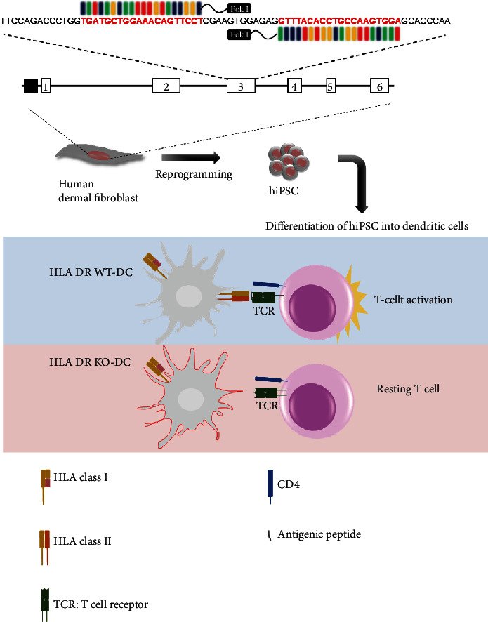
Figure 1Design of the TALEN for disrupting HLA DR expression on HDF and characterization of HLA DR knockout cells. (a) Schematic of TALEN binding sites within the HLA DR region. The red and underlined nucleotides highlight the anticipated binding sites for the left and right arms of TALEN. (b) Levels of HLA DR genetic disruption determined by the T7E1 assay. The lower bands (arrows) mark the digestion products, indicating TALEN-mediated gene modification. (c) Fluorescent intensity of wild-type and mutant fibroblast cells are different after transfection of the TALEN-encoding plasmids. (d) Alignment of the genomic sequences of wild type and mutant clone at the TALEN recognition site.Figure 2Loss of HLA DR expression on HLA DR knockout fibroblast after genome editing with TALENs. Loss of HLA DR protein expression in HLA DR knockout cells. HLA DR protein expression in wild-type and HLA DR knockout fibroblasts was analyzed by (a) FACS analysis and (b) western blotting. IFN-γ (50 ng/ml or 200 ng/ml) was treated for 5 days on both cell types.Figure 3Characterization of wild-type and HLA DR knockout iPSCs. Both wild-type and HLA DR knockout iPSCs show similar stemness potential. (a) Alkaline phosphatase staining of WT and HLA DR knockout iPS colonies. (b) Immunostaining of iPSCs for pluripotency-related genes Nanog, Oct4, and SSEA4. (c) Teratoma formation showed that wild-type and HLA DR knockout iPSCs differentiated into three germ layers of tissues in vivo (scale bars: 200 μm) and (d) compared the expression of HLA DR expression in CD45-positive tissues (scale bars: 200 μm). Only wild type teratomas showed HLA DR expression in CD45-positive tissues.Figure 4Differentiation of dendritic cells derived from wild-type and HLA DR knockout iPSCs. (a) Schematic representation of differentiation protocol for iPSCs into dendritic cells. (b) iPS cell-derived dendritic cells on day 4 in the 1st step, day 6 in the 2nd step, day 13 in the 3rd step, day 24 in the 4th step, and day 31 in the 5th step were analyzed for the expression of brachyury, CD34, CD45, CD14, and CD83, respectively. (c) May-Grunwald-Giemsa staining of mature iPS induced DC on the glass slide is shown.Figure 5Validation of HLA DR knockout iPSC immunogenicity. CD83-positive matured DC from HLA DR knockout iPSCs does not express HLA DR, and there is no response to the CD4 T cells. (a) FACS analysis showed the expression of CD83 and HLA DR for wild-type iPSC-derived DC vs. HLA DR knockout iPSC-derived DC. (b) MLR assay revealed that wild-type iPSC-derived DC significantly induced allogeneic lymphocyte activation compared with HLA DR knockout iPSC-derived DC. (The asterisks indicate statistically significant changes: ∗P ≤ 0.5, ∗∗P ≤ 0.01, N = 3).Figure 6Establishment of universally compatible immune nonresponsive human iPSCs by genome editing. Mismatches at the HLA DR (representative HLA class II) locus reveal the greatest impact on the development of an alloimmune response against transplanted organs such as the heart, kidney, and lung. Transcription activator-like effector nucleases (TALENs) were designed for selective removal of HLA DR expression. The TALENSs completely disrupted the expression of HLA DR on human dermal fibroblast cells (HDFs). Dendritic cells derived (differentiated) from HLA DR knockout iPSCs did not express HLA DR and reduced CD4+ T cell activation. These engineered iPSCs may have resolved the problem of immune rejection and provided a novel clinical application of derivatives.