SCI 발표논문

SCI 발표논문

← SCI 발표논문 목록

Stem Cells Int. 2017;2017:5180579.

Human Chorionic Plate-Derived Mesenchymal Stem Cells Restore Hepatic Lipid Metabolism in a Rat Model of Bile Duct Ligation.

, , , , , ,

제1저자 이윤빈, 최종호 · 교신저자 윤정환, 김기진

소속 (5)
  1. Department of Internal Medicine, CHA Bundang Medical Center, CHA University, Seongnam, Republic of Korea.
  2. Department of Biomedical Science, CHA University, Seongnam, Republic of Korea.
  3. Department of Dermatology, The Feinberg School of Medicine, Northwestern University, Chicago, IL, USA.
  4. Clinical Research Center, CHA Bundang Medical Center, CHA University, Seongnam, Republic of Korea.
  5. Department of Internal Medicine and Liver Research Institute, Seoul National University College of Medicine, Seoul, Republic of Korea.

Abstract

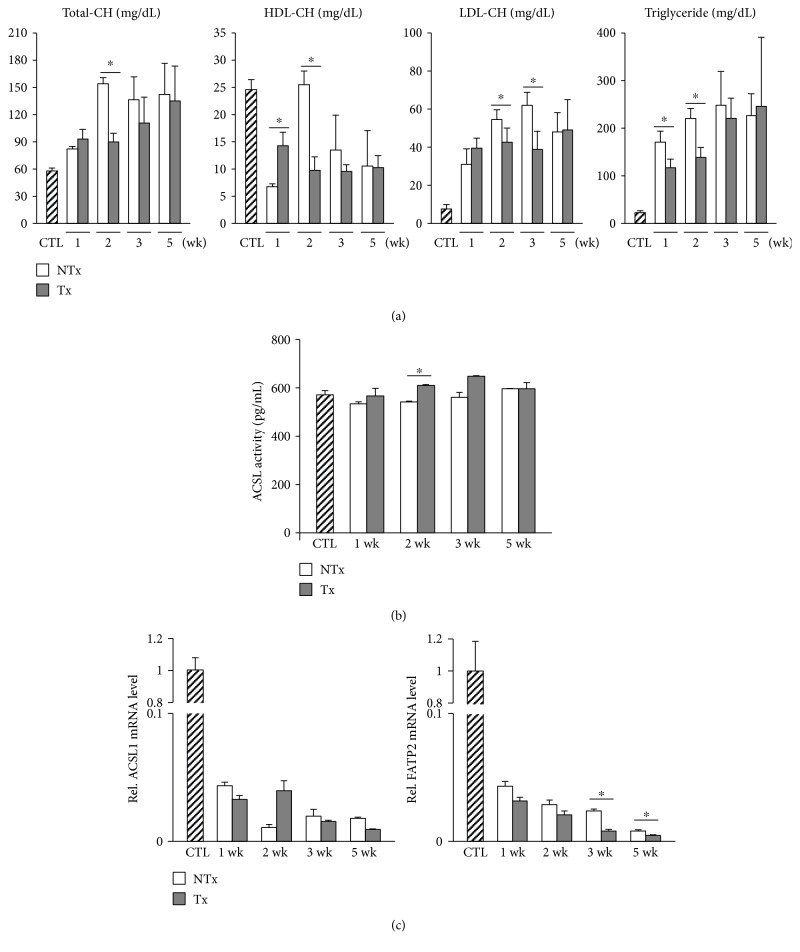
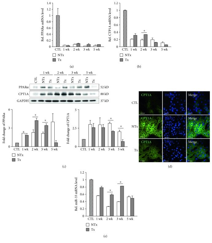
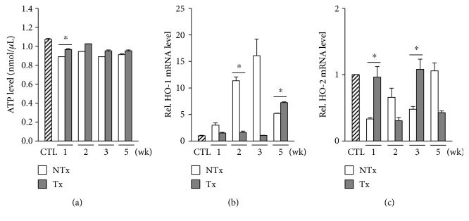
In cholestatic liver diseases, impaired bile excretion disrupts lipid homeostasis. We investigated changes of lipid metabolism, including mitochondrial β-oxidation, in a rat model of bile duct ligation (BDL) in which chorionic plate-derived mesenchymal stem cells (CP-MSCs) were transplanted. Serum cholesterol level, which was elevated after BDL, was significantly decreased following CP-MSC transplantation. The expression levels of genes involved in intracellular lipid uptake, including long-chain fatty acyl-CoA synthetases and fatty acid transport proteins, were decreased in rats after BDL; however, they were not significantly changed by subsequent CP-MSC transplantation. Carnitine palmitoyltransferase 1A (CPT1A), a rate-limiting enzyme in mitochondrial β-oxidation, was upregulated after BDL and then was downregulated after CP-MSC transplantation. CPT1A expression was changed via microRNA-33-a posttranscriptional regulator of CPT1A-in a peroxisome proliferator-activated receptor α-independent manner. Cellular adenosine triphosphate production-an indicator of mitochondrial function-was reduced after BDL and was restored by CP-MSC transplantation. Expression levels of heme oxygenases also were significantly affected following BDL and CP-MSC transplantation. Lipid metabolism is altered in response to chronic cholestatic liver injury and can be restored by CP-MSC transplantation. Our study findings support the therapeutic potential of CP-MSCs in cholestatic liver diseases and help in understanding the fundamental mechanisms by which CP-MSCs affect energy metabolism.

Figures
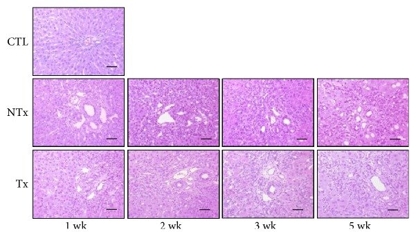
Figure 1
Figure 1 Inflammatory response induced by chronic cholestasis and the effect of CP-MSC transplantation. Histological analysis with hematoxylin and eosin staining (scale bar = 50 μm; original magnification, ×200). CTL: control group; NTx: nontransplanted group; Tx: transplanted group.
Figure 2
Figure 2 Changes in serum lipid profiles and expression levels of genes associated with intracellular uptake of fatty acids after BDL and/or CP-MSC transplantation. (a) Serum levels of total cholesterol, HDL cholesterol, LDL cholesterol, and triglyceride. (b) Activities of ACSL, as measured by ELISA. (c) mRNA expression levels of ACSL1 (left) and FATP2 (right). β-Actin was used as an internal control for normalization. Data are expressed as a fold change related to the control group. ∗P < 0.05 (compared to the nontransplanted group). CTL: control group; NTx: nontransplanted group; Tx: transplanted group.
Figure 3
Figure 3 Expression of genes associated with fatty acid oxidation after BDL and/or CP-MSC transplantation. mRNA expression levels of PPARα (a) and CPT1A (b) by real-time PCR. β-Actin was used as an internal control for normalization. Data are expressed as a fold change related to the control group. (c) Protein expression levels of PPARα and CPT1A. GAPDH was used as a loading control, and quantification by densitometry of Western blots was normalized to GAPDH. Data are expressed as a fold change related to the control group. (d) Analysis of CPT1A expression with immunofluorescence staining (scale bar = 200 μm; original magnification, ×400). Liver tissues, which were collected at 3 weeks posttransplantation in the transplanted group and post-BDL in the nontransplanted group, were used in immunofluorescence staining. (e) mRNA expression levels of miR-33. U6 snRNA was used as an internal control for normalization. ∗P < 0.05 (compared to the nontransplanted group). CTL: control group; NTx: nontransplanted group; Tx: transplanted group.
Figure 4
Figure 4 Changes in cellular ATP production and expression levels of HOs after BDL and/or CP-MSC transplantation. (a) Analysis of ATP levels in the liver tissues by the ATP assay. mRNA expression levels of HO-1 (b) and HO-2 (c), assessed by real-time PCR. GAPDH was used as an internal control for normalization. Data are expressed as a fold change related to the control group. ∗P < 0.05 (compared to the nontransplanted group). CTL: control group; NTx: nontransplanted group; Tx: transplanted group.

그림 출처: PubMed Central 공개 논문 (PMC5700509)

그 밖의 정보
출판 형태
Journal Article
저널
Stem cells international · ISSN 1687-966X
전자 출판
2017-11-09
참고문헌
27편