
BMC Gastroenterol. 2020;20(1):51.
Clinical significance of regional lymph node enlargement in patients with EGC within the expanded criteria for ESD.
제1저자 이동석 · 교신저자 박종규
소속 (3)
- Department of Gastroenterology, SMG-SNU Boramae Medical Center, Seoul National University of College of Medicine, Seoul, South Korea.
- Department of Gastroenterology, Gangneung Asan Hospital, University of Ulsan College of Medicine, Gangneung, South Korea. sajahoooo@naver.com.
- Department of Gastroenterology, Gangneung Asan Hospital, University of Ulsan College of Medicine, Gangneung, South Korea.
Abstract
BACKGROUND Lymph node (LN) metastasis is negligible in early gastric cancer (EGC) within expanded criteria for endoscopic submucosal dissection (ESD). However, regional lymph nodes in abdominal CT scans are sometimes enlarged in patients with EGC within the expanded criteria for endoscopic submucosal dissection (ESD). In this study, we investigated the clinical significance of regional lymph node enlargement on abdominal CT scan in patients with EGC within the expanded criteria for ESD.
METHODS From December 2010 to April 2015, among 301 patients with EGC within the ESD expanded criteria, 47 patients with regional lymph node enlargement shown by abdominal CT scan were prospectively enrolled. We performed surgical resection or periodic follow-up with abdominal CT scans and upper endoscopy every 6 months to evaluate whether the enlarged lymph nodes are due to metastasis or a reactive change.
RESULTS The mean age of the 47 patients (38 males, 9 female) was 64.8 years. The enlarged lymph nodes were usually single (26/47, 44.6%) and sized as follows: 11 nodes were ≤ 5 mm, 19 were 6-10 mm, and 17 were ≥ 10 mm. Four of the 47 patients initially underwent surgical resection, and 8 patients underwent surgical resection after ESD. However, there was no lymph node metastasis in surgical specimens. Thirty-five patients received ESD and periodically followed up at a median duration of 56 months (IQR: 44-59 month). The enlarged lymph node disappeared in 12 of 35 patients, decreased in 9 patients and remained the same size in 13 patients, and increased in 1 patient.
CONCLUSION Regional lymph node enlargement on abdominal CT scan in patients within expanded criteria for ESD of ECG may be not due to metastasis but a reactive change.
Keywords
Early gastric cancerEndoscopic resectionLymph node enlargement
Figures
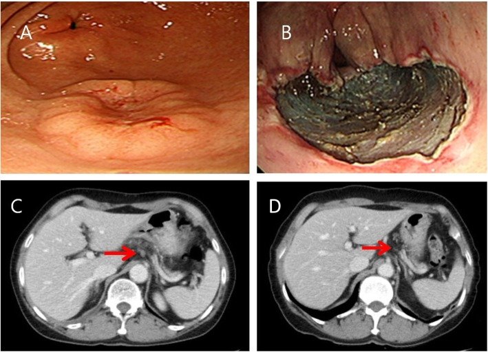
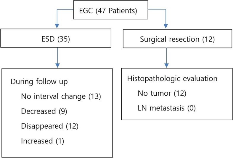
그림 출처: PubMed Central 공개 논문 (PMC7059300)
Conflict of interest statement
All authors have no actual or potential conflict of interest including any financial, personal or other relationships with other people or organizations.
그 밖의 정보
- 출판 형태
- Journal Article
- 저널
- BMC gastroenterology · eISSN 1471-230X
- 전자 출판
- 2020-03-05
- MeSH
- Aged · Early Detection of Cancer · Endoscopic Mucosal Resection* · Female · Gastroscopy · Humans · Lymph Nodes / pathology · Lymphatic Metastasis / pathology · Male · Middle Aged · Stomach Neoplasms / pathology / surgery
- 참고문헌
- 20편