Therapeutic effect of a novel histone deacetylase 6 inhibitor, CKD-L, on collagen-induced arthritis in vivo and regulatory T cells in rheumatoid arthritis in vitro.
Department of Molecular Medicine and Biopharmaceutical Sciences, BK 21 plus Graduate School of Convergence Science and Technology, College of Medicine, Seoul National University, Seoul, Korea.
Department of Pharmacology and Toxicology, CKD Research Institute, CKD Pharmaceutical Company, Seoul, Korea.
Division of Rheumatology, Department of Internal Medicine, Seoul National University College of Medicine, Seoul, Korea.
Department of Molecular Medicine and Biopharmaceutical Sciences, BK 21 plus Graduate School of Convergence Science and Technology, College of Medicine, Seoul National University, Seoul, Korea. ysong@snu.ac.kr.
Division of Rheumatology, Department of Internal Medicine, Seoul National University College of Medicine, Seoul, Korea. ysong@snu.ac.kr.
Abstract
BACKGROUND Histone deacetylase (HDAC) inhibitor has recently been reported to have a therapeutic effect as an anti-inflammatory agent in collagen-induced arthritis (CIA). We investigated the therapeutic effect of a new selective HDAC6 inhibitor, CKD-L, compared to ITF 2357 or Tubastatin A on CIA and regulatory T (Treg) cells in patients with rheumatoid arthritis (RA).
METHODS CIA was induced by bovine type II collagen (CII) in DBA/1 J mice. Mice were treated with HDAC inhibitor for 18 days. Arthritis score was assessed and histological analysis was performed by hematoxylin and eosin (H&E) stain. Cytotoxic T-lymphocyte associated protein (CTLA)-4 expression in induced Treg cells was analyzed and suppression assay was analyzed using Treg cells and effector T (Teff) cells isolated from naive C57BL/6 mice by flow cytometry. Cytokines were analyzed in peripheral blood mononuclear cells (PBMC) of five patients with RA by enzyme-linked immunosorbent assay (ELISA) and real-time polymerase chain reaction (PCR). Tumor necrosis factor (TNF) was analyzed using PMA- activated THP-1 cells by ELISA. Suppression assay was analyzed using Treg cells and Teff cells isolated from RA patients by flow cytometry.
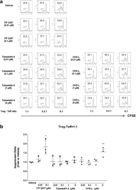
RESULTS In the CIA model, CKD-L and Tubastatin A significantly decreased the arthritis score. CKD-L increased CTLA-4 expression in Foxp3+ T cells and inhibited the proliferation of Teff cells in the suppression assay. In RA PBMC, CKD-L significantly inhibited TNF and interleukin (IL)-1β, and increased IL-10. CKD-L and Tubastatin A inhibited TNF secretion from PMA-activated THP-1 cells. CKD-L and ITF 2357 inhibited the proliferation of Teff cells in RA patients in the suppression assay. Tubastatin A had no effect on inhibition of proliferation.
CONCLUSION CKD-L decreased the arthritis score in CIA, reduced the expression of TNF and IL-1β, and increased the expression of IL-10 in PBMC from RA patients. CKD-L increased CTLA-4 expression and the suppressive function of Treg cells. These results suggest that CKD-L may have a beneficial effect in the treatment of RA.
Keywords
Collagen-induced arthritisHistone deacetylase 6Histone deacetylase inhibitorRegulatory T cellRheumatoid arthritis
Figures
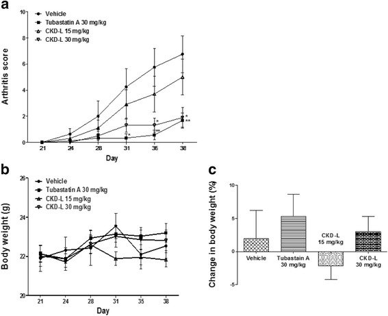
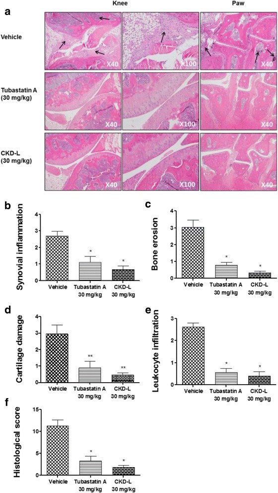
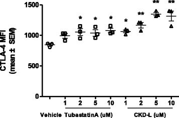
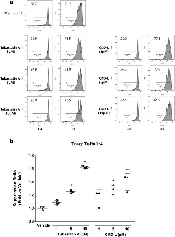
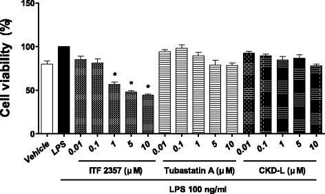
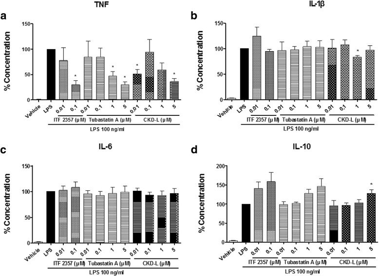
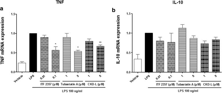
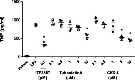
Fig. 1Therapeutic effects of CKD-L on CIA mice. After the onset of arthritis, mice were treated with vehicle, Tubastatin A (30 mg/kg), or CKD-L (15 or 30 mg/kg) subcutaneously every day for 18 days after the second immunization. CKD-L (30 mg/kg) and Tubastatin A significantly decreased arthritis score compared to vehicle (a). Body weight did not change during drug administration (b). The changes in body weight between day 21 and day 38 was not significantly different among the groups (c). *p < 0.05, **p < 0.01, vs vehicleFig. 2Histological analysis of CIA treated with CKD-L or Tubastatin A. Histological analyses were performed on knee joints (×40, ×100) and hind paws (×40) stained by H&E. Synovial inflammation, bone erosion, cartilage damage, and leukocyte infiltration were observed in the knee joints and hind paws of mice treated with vehicle (arrows) compared to Tubastatin A or CKD-L (a). The mean score of synovial inflammation (b), bone erosion (c), cartilage damage (d), and leukocyte infiltration (e) were calculated using a scale of 0–4 (normal = 0; mild = 1; moderate = 2; marked = 3; severe = 4), and these parameters significantly decreased after CKD-L or Tubastatin A treatment compared to vehicle treatment. Histological score was calculated by summation of these parameters (f). Histological score significantly decreased following treatment with CKD-L or Tubastatin A. *p < 0.001, **p < 0.01, vs vehicleFig. 3Increased cytotoxic T-lymphocyte antigen-4 (CTLA-4) expression by CKD-L in Treg cells from C57BL/6 mice (n = 3). CD4+CD25– T cells were isolated from splenocytes of C57BL/6 mice. CD4+CD25– T cells were incubated with vehicle or HDAC6 inhibitors (1 to 10 μM) in the presence of antiCD3/CD28 beads and recombinant TGF-β2 in a 48-well plate for 6 days. CTLA-4 expression in CD4+CD25+Foxp3+ T cells was analyzed by mean fluorescence intensity (MFI; mean ± SEM) using flow cytometry. *p < 0.05, **p < 0.001, vs vehicleFig. 4Effect of CKD-L in the suppression assay using regulatory T (Treg) cells and effector T (Teff) cells from C57BL/6 mice (n = 3). Treg cells were mixed with eFluor®670-labeled Teff cells at ratios of 4:1 and 0:1. The mixed cells were incubated with vehicle or HDAC6 inhibitor (1 to10 μM) in the presence of antiCD3/CD28 beads for 4 days. a The proliferation of Teff cells was assessed by eFluor®670 dilution using flow cytometry (representative data). b Suppression ratio (fold inhibition of cell proliferation by HDACi vs. vehicle) at a ratio of Treg:Teff = 1:4. *p < 0.01, **p < 0.001, vs vehicleFig. 5Effects of CKD-L on cell viability in PBMC from RA patients (n = 6). PBMC from RA patients were cultured in a 96-well plate for 24 h in the presence of lipopolysaccharide (LPS; 100 ng/ml) and HDAC inhibitors at different concentrations (0.01 to 5 μM). Cell viability was determined using the MTT assay. Bars represent means and SDs. All experiments were carried out in triplicate. *p < 0.001, vs LPSFig. 6Effects of CKD-L on cytokine secretion in PBMC from RA patients (n = 5). PBMC from RA patients were cultured in a 48-well plate for 24 h in the presence of lipopolysaccharide (LPS; 100 ng/ml) and HDAC inhibitors at different concentrations (0.01 to 5 μM). Tumor necrosis factor (TNF) (a), IL-1β (b), IL-6 (c), and IL-10 (d) were measured in the culture supernatant by ELISA. *p < 0.01, vs LPS. IL interleukinFig. 7Effects of CKD-L on cytokine mRNA expression in PBMC from RA patients (n = 3). PBMC from RA patients were cultured in a 48-well plate for 24 h in the presence of lipopolysaccharide (LPS; 100 ng/ml) and HDAC inhibitors at different concentrations (0.01 to 5 μM). Total RNA was extracted from harvested cells and cDNA was synthesized by RT-PCR. Tumor necrosis factor (TNF) (a) and interleukin-10 (IL-10) (b) mRNA expression levels were analyzed by real-time PCR. *p < 0.001, **p < 0.01, vs LPSFig. 8Tumor necrosis factor (TNF) secretion in PMA-activated THP-1 cells (n = 3). THP-1 cells (1 × 10⁵ cells/well) were cultured in a 24-well plate and activated with PMA (10 ng/ml) for 24 h to induce macrophage differentiation. PMA-activated THP-1 cells were treated with vehicle or HDAC inhibitors (0.1 to 10 μM) for 24 h and then treated with 100 ng/ml lipopolysaccharide (LPS) for 4 h. The secretion of TNF was measured by ELISA. *p < 0.001, vs LPSFig. 9Effects of CKD-L in the suppression assay using induced regulatory T (Treg) cells and effector T (Teff) cells from RA patients (n = 3). Treg cells induced from RA patients were mixed with carboxyfluorescein succinimidyl ester (CFSE)-labeled Teff cells at ratios of 1:1, 0.3:1, and 0:1. The mixed cells were incubated with vehicle or HDAC inhibitors (0.01 to 5 μM) in the presence of Dynabeads® human T-activator CD3/C28 (one bead to four cells) for 3 days. a The proliferation of Teff cells was assessed by CFSE dilution using flow cytometry (representative data). b Suppression ratio (fold inhibition of cell proliferation by HDACi vs vehicle) at the ratio of Treg:Teff = 1:1. *p <0.001, **p < 0.05, vs vehicle