
BMC Cancer. 2019;19(1):19.
Differences in tumor microenvironments between primary lung tumors and brain metastases in lung cancer patients: therapeutic implications for immune checkpoint inhibitors.
제1저자 김률 · 교신저자 김범석
소속 (7)
- Department of Internal Medicine, Seoul National University Hospital, 101 Daehak-ro, Jongno-gu, Seoul, 03080, Republic of Korea.
- Department of Internal Medicine, Seoul National University Hospital, 101 Daehak-ro, Jongno-gu, Seoul, 03080, Republic of Korea. bhumsuk@snu.ac.kr.
- Cancer Research Institute, Seoul National University College of Medicine, 101 Daehak-ro, Jongno-gu, Seoul, 03080, South Korea. bhumsuk@snu.ac.kr.
- Department of Pathology, Seoul National University Hospital, 101 Daehak-ro, Jongno-gu, Seoul, 03080, South Korea.
- Department of Internal Medicine, Seoul National University Bundang Hospital, 82 Gumi-ro 173 Beon-gil, Bundang-gu, Seongnam-si, Gyeonggi-do, 13620, South Korea.
- Department of Neurosurgery, Seoul National University Hospital, 101 Daehak-ro, Jongno-gu, Seoul, 03080, South Korea.
- Cancer Research Institute, Seoul National University College of Medicine, 101 Daehak-ro, Jongno-gu, Seoul, 03080, South Korea.
Abstract
BACKGROUND We aimed to compare intra- and extracranial responses to immune checkpoint inhibitors (ICIs) in lung cancer with brain metastases (BM), and to explore tumor microenvironments of the brain and lungs focusing on the programmed cell death-1 (PD-1)/programmed cell death ligand-1 (PD-L1) pathway.
METHODS Two cohorts of lung cancer patients with BM were analyzed. Cohort 1 included 18 patients treated with nivolumab or pembrolizumab, and intra- and extracranial responses were assessed. Cohort 2 comprised 20 patients who underwent both primary lung surgery and brain metastasectomy. Specimens from cohort 2 were subjected to immunohistochemical analysis for the following markers: CD3, CD4, CD8, FOXP3, and PD-1 on tumor infiltrating lymphocytes (TIL) and PD-L1 on tumor cells.
RESULTS Seven patients (38.9%) in cohort 1 showed progressive disease in both primary and intracranial lesions. Although the other 11 patients exhibited a partial response or stable disease in the primary lesion, eight showed a progression in BM. Interestingly, PD-1+ TILs were significantly decreased in BM (P = 0.034). For fifteen patients with adenocarcinoma, more distinctive patterns were observed in CD3+ (P = 0.078), CD8+ (P = 0.055), FOXP3+ (P = 0.016), and PD-1+ (P = 0.016) TILs.
CONCLUSIONS There may be discordant responses to an ICI of lung cancer between primary lung lesion and BM based on discrepancies in the tumor microenvironment. The diminished infiltration of PD-1+ TILs in tumor tissue within the brain may be one of the major factors that hinder the response to anti-PD-1 antibody in BM.
Keywords
Brain metastasisImmunotherapyLung cancerPD-1PD-L1
Figures
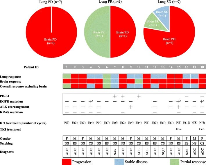
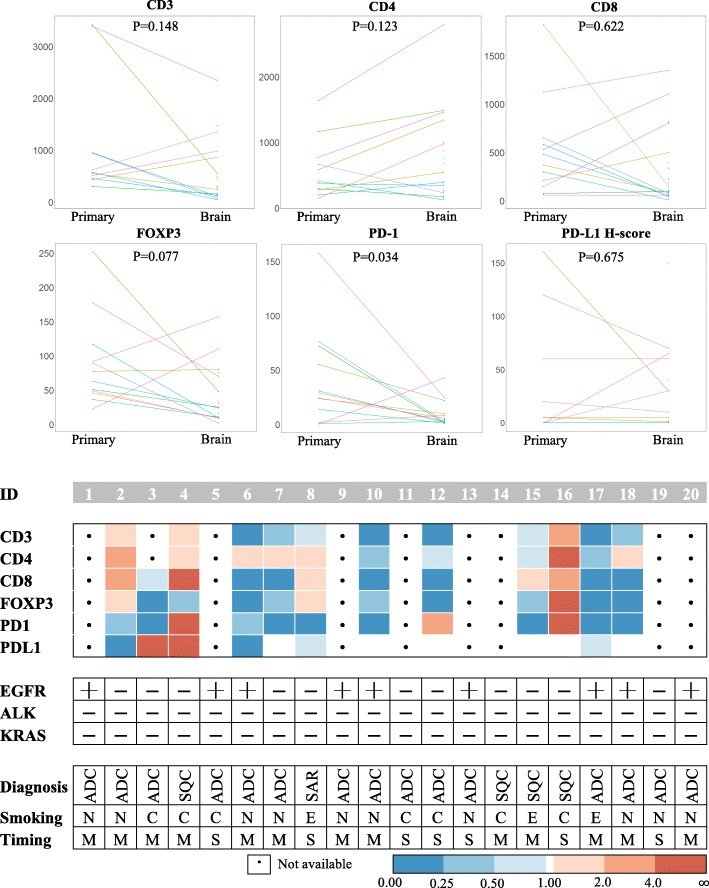
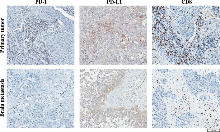
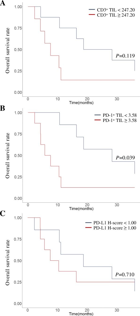
그림 출처: PubMed Central 공개 논문 (PMC6322302)
Conflict of interest statement
ETHICS APPROVAL AND CONSENT TO PARTICIPATE: This study was approved by the SNUH Institutional Review Board (IRB approval number: H-1702-158-836) and was conducted in accordance with Declaration of Helsinki provisions. Written informed consent to participate in the study was obtained from each patient at admission. CONSENT FOR PUBLICATION: Not applicable. COMPETING INTERESTS: The authors declare that they have no competing interests. PUBLISHER’S NOTE: Springer Nature remains neutral with regard to jurisdictional claims in published maps and institutional affiliations.
그 밖의 정보
- 출판 형태
- Journal Article
- 저널
- BMC cancer · eISSN 1471-2407
- 전자 출판
- 2019-01-07
- MeSH
- Adult · Aged · Antibodies, Monoclonal, Humanized / therapeutic use · Antineoplastic Agents, Immunological / therapeutic use · B7-H1 Antigen / immunology · Biomarkers, Tumor / immunology · Brain Neoplasms / drug therapy / immunology / secondary · Cohort Studies · Female · Humans · Immunomodulation · Lung Neoplasms / drug therapy / immunology / pathology · Lymphocytes, Tumor-Infiltrating / immunology · Male · Middle Aged · Nivolumab / therapeutic use · Programmed Cell Death 1 Receptor / immunology · Tumor Microenvironment* / immunology
- Substances
- Antibodies, Monoclonal, Humanized · Antineoplastic Agents, Immunological · B7-H1 Antigen · Biomarkers, Tumor · Programmed Cell Death 1 Receptor · Nivolumab · pembrolizumab
- 연구비
- Seoul National University Hospital (KR) 03-2015-0380
- 참고문헌
- 35편