Medical Science Research Institute, School of Medicine, Sungkyunkwan University (SKKU), Suwon 16419, Republic of Korea.
Department of Environmental Health Sciences, Graduate School of Public Health, Seoul National University, Seoul 08826, Republic of Korea.
Institute of Health and Environment, Seoul National University, Seoul 08826, Republic of Korea.
Division of Gastroenterology and Hepatology, Department of Internal Medicine, Seoul National University College of Medicine, Seoul Metropolitan Government Boramae Medical Center, Seoul 07061, Republic of Korea.
Department of Internal Medicine, Chungnam National University School of Medicine, Daejeon 35015, Republic of Korea.
Center for Human and Environmental Microbiome, Institute of Health and Environment, Seoul National University, Seoul 08826, Republic of Korea.
KoBioLabs, Inc., Seoul 08826, Republic of Korea.
Bio-MAX/N-Bio, Seoul National University, Seoul 08826, Republic of Korea.
Abstract
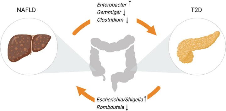
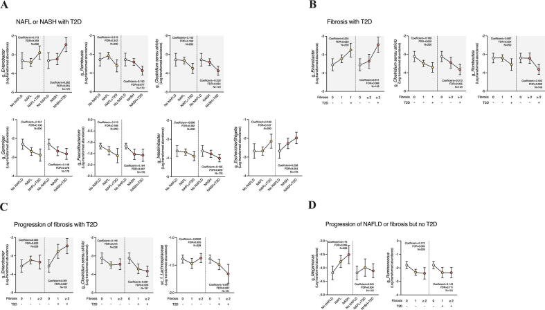
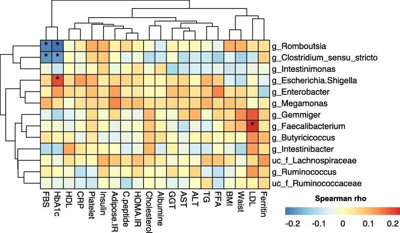
Non-alcoholic fatty liver disease (NAFLD) is closely associated with type 2 diabetes mellitus (T2D), and these two metabolic diseases demonstrate bidirectional influences. The identification of microbiome profiles that are specific to liver injury or impaired glucose metabolism may assist understanding of the role of the gut microbiota in the relationship between NAFLD and T2D. Here, we studied a biopsy-proven Asian NAFLD cohort (n = 329; 187 participants with NAFLD, 101 with NAFLD and T2D, and 41 with neither) and identified Enterobacter, Romboutsia, and Clostridium sensu stricto as the principal taxa associated with the severity of NAFLD and T2D, whereas Ruminococcus and Megamonas were specific to NAFLD. In particular, the taxa that were associated with both severe liver pathology and T2D were also significantly associated with markers of diabetes, such as fasting blood glucose and Hb1Ac. Enterotype analysis demonstrated that participants with NAFLD had a significantly higher proportion of Bacteroides and a lower proportion of Ruminococcus than a Korean healthy twin cohort (n = 756). However, T2D could not be clearly distinguished from NAFLD. Analysis of an independent T2D cohort (n = 185) permitted us to validate the T2D-specific bacterial signature identified in the NAFLD cohort. Functional inference analysis revealed that endotoxin biosynthesis pathways were significantly enriched in participants with NAFLD and T2D, compared with those with NAFLD alone. These findings may assist with the development of effective therapeutic approaches for metabolic diseases that are associated with specific bacterial signatures.
ALT, alanine aminotransferaseBMI, body mass indexBiomarkerEnterotypeFBS, fasting blood sugarFDR, false discovery rateFLI, fatty liver indexGut microbiomeHbA1c, glycosylated hemoglobinLDL, low-density lipoproteinLPS, lipopolysaccharideMaAsLin2, microbiome multivariable association with linear models 2NAFL, non-alcoholic fatty liverNAFLD, non-alcoholic fatty liver diseaseNASH, non-alcoholic steatohepatitisNASH-CRN, non-alcoholic steatohepatitis clinical research networkNon-alcoholic fatty liver disease (NAFLD)PICRUSt2, phylogenetic investigation of communities by reconstruction of unobserved states 2T2D, type 2 diabetes mellitusType 2 diabetes mellitus
Figures
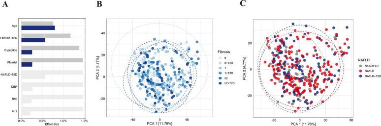
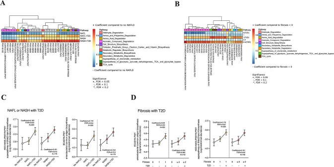
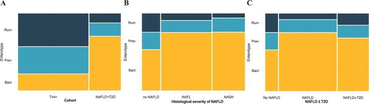
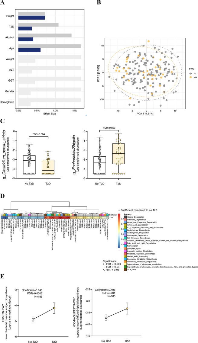
Fig. 1Variations in the microbial community in the NAFLD-T2D cohort. (A) Explanatory power of the characteristics of the cohort for the variations in the microbial community at the genus level. Blue and dark gray bars indicate the individual and cumulative effect sizes. Light gray bars indicate variables that were significant on an individual basis but were not included in the forward stepwise RDA model. Principal component analysis (PCA) was performed, based on Aitchison transformation, for (B) fibrosis and (C) NAFLD. n = 329; FDR < 0.1. (For interpretation of the references to color in this figure legend, the reader is referred to the web version of this article.)Fig. 2Comparisons of the abundances of microbial taxa in the NAFLD cohort, categorized according to either the histology of liver biopsies from participants with NAFLD and T2D or the severity of hepatic fibrosis in participants with T2D. The genera that were significantly associated with NAFLD are depicted as mean connected plots. Significant associations were found for (A) NAFL or NASH with T2D, (B) fibrosis with T2D only, (C) the progression of fibrosis with T2D, and (D) the progression of NAFLD or fibrosis but no T2D. Features with the background color indicate the overlapping features between the groups. Associations were identified using the multivariate association analysis, after adjustment for age, and the corrected significance levels were calculated using the Benjamini-Hochberg method (FDR < 0.2). n = 329.Fig. 3Relationships between the abundances of disease-associated taxa and host parameters. Correlation coefficients (adjusted for age) for the relationships between the relative abundances of 10 disease-associated taxa and 20 host parameters were calculated using Spearman’s correlation method. The color of the plot denotes the magnitude of Spearman’s rho (n = 329, * FDR < 0.1). Abbreviations: FBS, fasting blood sugar; HbA1c, glycosylated hemoglobin; HDL, high-density lipoprotein; CRP, high-sensitivity C-reactive protein; Adipose.IR, adipose tissue insulin resistance; C.peptide, connecting peptide; HOMA.IR, homeostasis model assessment of insulin resistance; GGT, gamma-glutamyl transferase; AST, aspartate aminotransferase; ALT, alanine aminotransferase; TG, triglycerides; FFA, free fatty acid; BMI, body mass index; Waist, waist circumference; LDL, low-density lipoprotein.Fig. 4Inferred microbial metabolic pathways in participants with NAFLD, categorized according to either the histology of a liver biopsy from participants with NAFLD and T2D or the severity of fibrosis in participants with T2D. The activity of metabolic pathways in the microbial community was predicted using PICRUSt2 and annotated using the MetaCyc database. (A) Thirty-one metabolic pathways were significantly upregulated in participants with NASH ± T2D vs. those without NAFLD, as shown in a heatmap. (B) Twenty-five metabolic pathways were upregulated in participants with a fibrosis score ≥ 2 ± T2D. The color of the plots denotes the magnitudes of the coefficients. The associations of T2D with two metabolic pathways related to lipopolysaccharide synthesis are also shown according to (C) the liver histology of participants with NAFLD and (D) the severity of hepatic fibrosis. The data were analyzed using the multivariate association analysis method, after adjustment for age, and FDR was calculated using the Benjamini-Hochberg method. n = 329; +, FDR < 0.05; −, FDR < 0.10; *FDR < 0.20.Fig. 5Enterotypes of the Korean population cohorts. (A) Proportions of each enterotype in the healthy Korean twin (n = 752) and NAFLD-T2D (n = 329) cohorts. (B) Proportions of each enterotype in the NAFLD-T2D cohort, categorized according to the presence of NAFLD alone (n = 228) and (C) NAFLD plus T2D (n = 329). FDR < 0.1.Fig. 6Validation of the identified NAFLD-T2D-associated bacterial signatures using an independent T2D cohort. (A) The explanatory power of the cohort variables for the variations in the microbial communities at the genus level. Blue and dark gray bars indicate individual and cumulative effect sizes, respectively. Light gray bars indicate variables that were significant on an individual basis but were not included in the forward stepwise RDA model. (B) Principal components analysis (PCA), based on Aitchison transformation, for T2D. (C) Differences in the abundance of bacterial taxa in participants with and without T2D in the validation cohort. (D) Forty-seven inferred metabolic pathways that were significantly upregulated in participants with T2D. (E) Comparisons of the pathways related to LPS biosynthesis. n = 185; FDR < 0.1. (For interpretation of the references to color in this figure legend, the reader is referred to the web version of this article.)
그림 출처: PubMed Central 공개 논문 (PMC8591343)
Conflict of interest statement
GK is a founder of KoBioLabs, Inc., a company that aims to characterize the role of host–microbiome interactions in chronic diseases. The other authors declare no competing interests.
그 밖의 정보
출판 형태
Journal Article
저널
Computational and structural biotechnology journal · ISSN 2001-0370