
BMC Musculoskelet Disord. 2016;17:42.
Femoral geometry, bone mineral density, and the risk of hip fracture in premenopausal women: a case control study.
제1저자 이동화 · 교신저자 김상완
소속 (8)
- Department of Internal Medicine, Seoul National University Hospital, 101 Daehak-ro, Jongno-gu, Seoul, 110-744, Korea. roroko0902@gmail.com.
- Department of Internal Medicine, Seoul National University Bundang Hospital, 300 Gumi-dong, Bundang-gu, Seongnam-si, Kyunggi-do, 463-707, Korea. yeun6486@gmail.com.
- Department of Internal Medicine, Seoul National University Hospital, 101 Daehak-ro, Jongno-gu, Seoul, 110-744, Korea. wanilove23@nate.com.
- Department of Internal Medicine, Seoul National University Hospital, 101 Daehak-ro, Jongno-gu, Seoul, 110-744, Korea. jhkxingfu@gmail.com.
- Department of Internal Medicine, Seoul National University Bundang Hospital, 300 Gumi-dong, Bundang-gu, Seongnam-si, Kyunggi-do, 463-707, Korea. kyoungmin02@gmail.com.
- Department of Internal Medicine, Seoul National University Hospital, 101 Daehak-ro, Jongno-gu, Seoul, 110-744, Korea. csshin@snu.ac.kr.
- Department of Internal Medicine, Seoul National University Hospital, 101 Daehak-ro, Jongno-gu, Seoul, 110-744, Korea. seongyk@plaza.snu.ac.kr.
- Department of Internal Medicine, Seoul Metropolitan Government Boramae Medical Center, 20 Boramae-Ro 5-Gil, Dongjak-Gu, Seoul, 156-707, Korea. swkimmd@snu.ac.kr.
Abstract
BACKGROUND The purpose of this study was to determine the relationships among hip geometry, bone mineral density, and the risk of hip fracture in premenopausal women.
METHODS The participants in this case-control study were 16 premenopausal women with minimal-trauma hip fractures (fracture group) and 80 age-and BMI-adjusted controls. Subjects underwent dual-energy X-ray absorptiometry (DXA) to assess BMD at the proximal femur and to obtain DXA-derived hip geometry measurements.
RESULTS The fracture group had a lower mean femoral neck and total hip BMD than the control group (0.721 ± 0.123 vs. 0.899 ± 0.115, p <0.001 for the femoral neck BMD and 0.724 ± 0.120 vs. 0.923 ± 0.116, p <0.001 for the total hip BMD). In addition, participants in the fracture group had a longer hip axis length (HAL; p = 0.007), narrower neck shaft angle (NSA; p = 0.008), smaller cross sectional area (CSA; p < 0.001) and higher cross sectional moment of inertia (CSMI; p = 0.004) than those in control group. After adjusting for BMD, the fracture group still had a significantly longer mean HAL (p = 0.020) and narrower NSA (p = 0.006) than the control group.
CONCLUSIONS BMD is an important predictor of hip fracture in premenopausal women. Furthermore, HAL and NSA are BMD-independent predictors of hip fracture in premenopausal women. Hip geometry may be clinically useful for identification of premenopausal women for whom active fracture prevention should be considered.
Figures
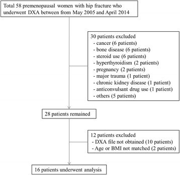
그림 출처: PubMed Central 공개 논문 (PMC4727305)
그 밖의 정보
- 출판 형태
- Journal Article
- 저널
- BMC musculoskeletal disorders · eISSN 1471-2474
- 전자 출판
- 2016-01-25
- MeSH
- Adult · Bone Density / physiology · Case-Control Studies · Female · Femur / diagnostic imaging · Hip Fractures / diagnostic imaging · Humans · Premenopause / physiology · Radiography · Retrospective Studies · Risk Factors
- 참고문헌
- 31편