Strategic Center of Cell & Bio Therapy, Seoul National University Hospital, Seoul 03080, Republic of Korea; Molecular Medicine and Biopharmaceutical Sciences, Graduate School of Convergence Science and Technology, Seoul National University, Seoul, Republic of Korea.
Department of Internal Medicine, Seoul National University Hospital, Seoul, Republic of Korea.
Strategic Center of Cell & Bio Therapy, Seoul National University Hospital, Seoul 03080, Republic of Korea; Program in Stem Cell Biology, Seoul National University College of Medicine, Seoul, Republic of Korea.
Strategic Center of Cell & Bio Therapy, Seoul National University Hospital, Seoul 03080, Republic of Korea; Molecular Medicine and Biopharmaceutical Sciences, Graduate School of Convergence Science and Technology, Seoul National University, Seoul, Republic of Korea; Program in Stem Cell Biology, Seoul National University College of Medicine, Seoul, Republic of Korea. Electronic address: hyosoo@snu.ac.kr.
Abstract
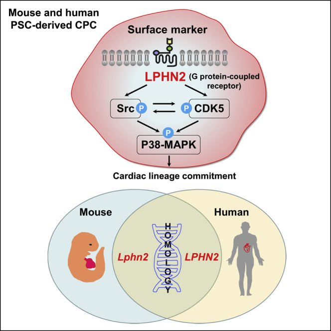
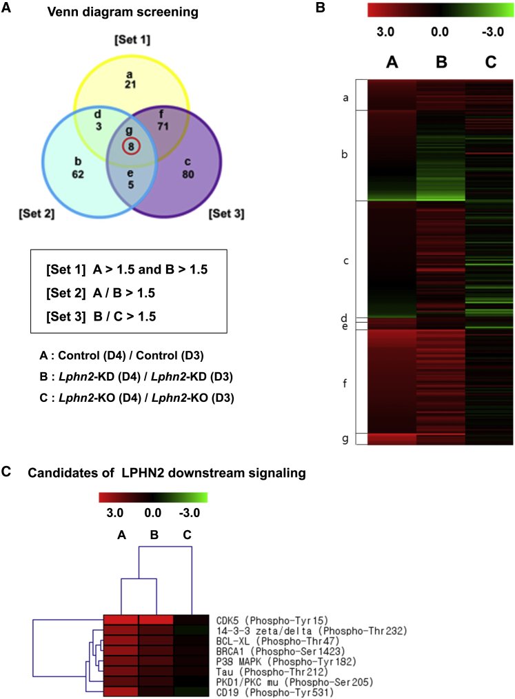
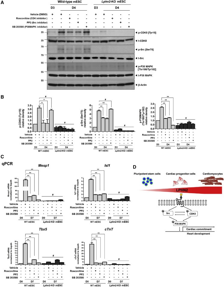
Identifying lineage-specific markers is pivotal for understanding developmental processes and developing cell therapies. Here, we investigated the functioning of a cardiomyogenic cell-surface marker, latrophilin-2 (LPHN2), an adhesion G-protein-coupled receptor, in cardiac differentiation. LPHN2 was selectively expressed in cardiac progenitor cells (CPCs) and cardiomyocytes (CMCs) during mouse and human pluripotent stem cell (PSC) differentiation; cell sorting with an anti-LPHN2 antibody promoted the isolation of populations highly enriched in CPCs and CMCs. Lphn2 knockdown or knockout PSCs did not express cardiac genes. We used the Phospho Explorer Antibody Array, which encompasses nearly all known signaling pathways, to assess molecular mechanisms underlying LPHN2-induced cardiac differentiation. LPHN2-dependent phosphorylation was the strongest for cyclin-dependent kinase 5 (CDK5) at Tyr15. We identified CDK5, Src, and P38MAPK as key downstream molecules of LPHN2 signaling. These findings provide a valuable strategy for isolating CPCs and CMCs from PSCs and insights into the still-unknown cardiac differentiation mechanisms.
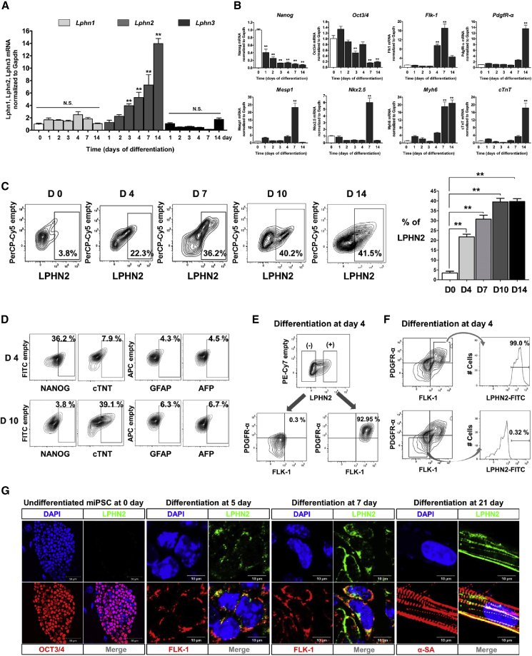
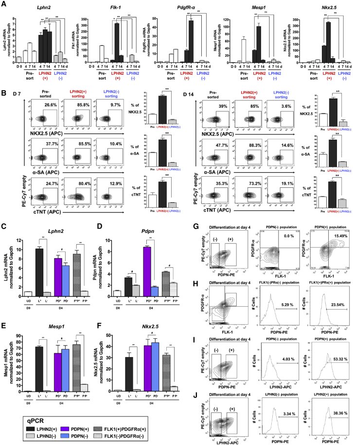
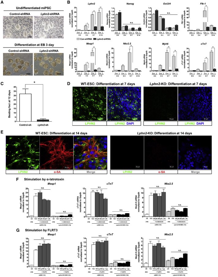
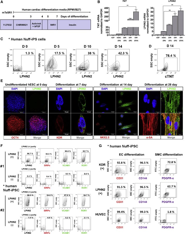
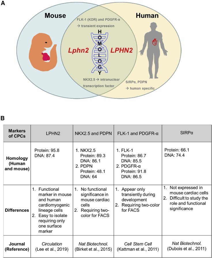
Figure 1Expression Pattern of LPHN2 during Cardiac Differentiation (A) Gene expression analysis of the latrophilin family in mouse induced pluripotent stem cells (iPSCs) during serial differentiation stages. Cultures of iPSCs under differentiation were harvested at the indicated days, and gene expression in cells was analyzed by qPCR. Values are shown relative to day 0. ∗∗p < 0.01, N.S., not significant, one-way ANOVA and post hoc Bonferroni test, n = 3 independent replicates. (B) mRNA expression levels of Nanog, Oct3/4, Flk-1, PdgfR-α, Mesp1, Nkx2.5, Myh6, and cTnT in mouse iPSC-derived cells at successive stages during cardiac differentiation. ∗∗p < 0.01, one-way ANOVA and post hoc Bonferroni test, n = 3 independent replicates. (C) Flow cytometric analysis of LPHN2 expression during cardiac differentiation. (Left) LPHN2 expression in mouse iPSC-derived cardiac cells at various time points. (Right) Quantification of the plots shown on the left. ∗∗p < 0.01, one-way ANOVA and post hoc Bonferroni test, n = 4 independent replicates. (D) Flow cytometric analysis of cells on consecutive days after cardiac differentiation. NANOG was used as a pluripotency marker, cTNT as a CMC marker, glial fibrillary acidic protein (GFAP) as an ectoderm marker, and α-fetoprotein (AFP) as an endoderm marker. (E and F) Representative fluorescence-activated cell sorting (FACS) plots showing the correlation between LPHN2 and co-expression of FLK-1 and PDGFR-α. FACS analysis for LPHN2 expression in differentiated cells at day 4 is shown. (E) The LPHN2+ and LPHN2− cell populations were analyzed using anti-FLK-1 and anti-PDGFR-α antibodies. (F) The FLK-1+/PDGFR-α+ and FLK-1−/PDGFR-α− cell populations were divided, and LPHN2 expression was analyzed. (G) Immunostaining for LPHN2 and OCT3/4, FLK-1, or α-SA in monolayer cultures in the early (day 7) and late (day 21) stages of cardiac differentiation. Blue, nuclear counterstaining with 4,6-diamidino-2-phenylindole (DAPI). PSC, pluripotent stem cell.Figure 2Enrichment of iPSC-Derived Cardiac Progenitor Cells and Cardiomyocytes by Cell Sorting Based on LPHN2 Expression (A) qPCR analysis of cardiac lineage cell markers in pre-sorting and sorted (LPHN2+ and LPHN2−) cells at various stages of cardiac differentiation of iPSCs. Values are shown relative to day 0. ∗p < 0.05, ∗∗p < 0.01, one-way ANOVA and post hoc Bonferroni test; n = 3 independent replicates. (B) Pre-sorting and sorted (LPHN2+ and LPHN2−) fractions at each time point were analyzed for NKX2.5, α-SA, and cTNT expression by intracellular flow cytometry. Quantification of the plots is shown on the right. ∗∗p < 0.01, one-way ANOVA and post hoc Bonferroni test; n = 3 independent replicates. (C and D) qPCR analysis of (C) Lphn2 and (D) Pdpn expression in the sorted cell populations on day 4 of differentiation. ∗∗p < 0.01, #not significant, one-way ANOVA and post hoc Bonferroni test; n = 3 independent replicates. UD, undifferentiated. (E and F) Differences in the mRNA expression of (E) Mesp1 and (F) Nkx2.5 in the sorted cell populations on day 4 of differentiation. Values are shown relative to day 0. ∗∗p < 0.01, #not significant, one-way ANOVA and post hoc Bonferroni test; n = 3 independent replicates. UD, undifferentiated. (G and H) Representative flow cytometric plots showing correlation in the expression of the cardiac progenitor cell markers PDPN, FLK-1, and PDGFR-α on day 4 of differentiation. (I and J) Representative flow cytometric plots showing correlation between LPHN2 and PDPN expression on day 4 of differentiation.Figure 3Functional Significance of LPHN2 during In Vitro Cardiac Differentiation (A) Gross morphology of control induced pluripotent stem cells (iPSCs) and Lphn2-knockdown (KD) iPSCs transduced by control-shRNA and Lphn2-shRNA, respectively. No differences were observed in the colony formation or EB formation. Scale bar, 200 μm. EB, embryoid body. (B) Effects of Lphn2 KD on cardiac differentiation. Transcriptional profiling of Lphn2, Nanog, Oct3/4, Flk-1, Mesp1, Nkx2.5, Myh6, and cTnT in control iPSC- and Lphn2-KD iPSC-derived cells by qPCR. ∗p < 0.05, ∗∗p < 0.01, N.S., not significant, one-way ANOVA and post hoc Bonferroni test; n = 3 independent replicates. (C) Quantification of spontaneous beating foci in control and Lphn2-KD cells on day 12 of cardiac differentiation. ∗p < 0.05, Mann-Whitney U test; n = 4 independent replicates. (D) Immunostaining for LPHN2 (green) expression in wild-type embryonic stem cell (WT-ESC) and Lphn2-knockout (KO) ESC-derived cells on day 7 of cardiac differentiation. Blue, nuclear counterstaining with DAPI. Scale bar, 50 μm. (E) WT-ESCs express LPHN2 (green) and α-SA (red) with striations at day 14 after differentiation, indicating cardiomyocyte differentiation, but Lphn2-KO ESCs did not differentiate into cardiomyocytes. Blue, nuclear counterstaining with DAPI. Scale bar, 20 μm. (F and G) qPCR analysis of gene expression of cardiac-specific Mesp1, Nkx2.5, and cTnT after stimulation with presumptive ligands. Treatment with α-latrotoxin (F) and FLRT3 (G) did not increase the expression of cardiac lineage genes in mESCs or Lphn2-KO ESCs after 7 days under cardiac differentiation conditions. Values are shown relative to the expression in WT-mESCs on day 0. N.S., not significant, one-way ANOVA and post hoc Bonferroni test; n = 3 independent replicates. UD, undifferentiated; Veh, vehicle. See also Videos S1, S2, S3, and S4.Figure 4Screening of LPHN2-mediated Cardiac Differentiation Mechanisms Based on the Phospho Explorer Antibody Array (A) A Venn diagram showing the intersection of sets 1, 2, and 3, with the most highly phosphorylated proteins in group A during differentiation of control-shRNA pluripotent stem cells. Lphn2-KO, Lphn2-knockout; Lphn2-KD, Lphn2-knockdown. (B) Heatmap constructed from the data shown in (A) shows the differential phosphorylation ratios in groups A, B, and C. (C) Phosphorylation sites of the most highly phosphorylated proteins in group A were considered as candidates for LPHN2 downstream signaling.Figure 5CDK5 Activated P38MAPK via Src and Induced Differentiation of Pluripotent Stem Cells into Cardiomyocytes (A) Phosphorylation levels of CDK5, Src, and P38MAPK in wild-type mouse ESCs (mESCs) and Lphn2-knockout (KO) mESCs treated with the respective inhibitors during cardiac differentiation, as detected by western blotting. (B) Quantification of the phosphorylation ratios of CDK5, Src, and P38MAPK in wild-type mESCs (WT mESCs) and Lphn2-KO mESCs. Values are shown relative to the phosphorylation ratio for the vehicle group of WT mESCs on day 3 of differentiation. ∗p < 0.05, ∗∗p < 0.01, #not significant, one-way ANOVA and post hoc Bonferroni test; n = 3 independent replicates. (C) Gene expression analysis of Mesp1, Isl1, Tbx5, and cTnT in WT mESCs and Lphn2-KO mESCs treated with inhibitors of CDK5, Src, and P38MAPK on day 7 of cardiac differentiation. Values are shown relative to the expression in WT mESCs on day 0. ∗∗p < 0.01, #not significant, one-way ANOVA and post hoc Bonferroni test; n = 3 independent replicates. (D) Schematic illustration of the molecular mechanism of LPHN2-mediated cardiac differentiation via the CDK5-Src-P38MAPK signaling cascade.Figure 6LPHN2 Expression and Enriched CPC Potential in Human Pluripotent Stem Cells (A) A schematic timeline demonstrating the major steps of the culturing procedure used for the directed differentiation of human pluripotent stem cells into cardiomyocytes. (B) qPCR analysis of TNT and LPHN2 expression in human embryonic stem cell (ESC)-derived cardiomyocytes. Expression values are shown relative to that in hDFs. ∗∗p < 0.01, one-way ANOVA and post hoc Bonferroni test; n = 3 independent replicates. hDF, human dermal fibroblast cell; hESC, human embryonic stem cell; Diff, differentiated. (C) FACS plots for LPHN2 expression in undifferentiated human Nuff-iPS cells and during a time course of cardiac differentiation. (D) FACS analysis of the cTNT expression in human Nuff-iPS cells during cardiac differentiation at day 14. (E) Immunostaining for LPHN2 (green), OCT4 (red), KDR (red), NKX2.5 (red), and α-SA (red) expression in human ESCs and ESC-derived cardiomyocytes. Blue, nuclear counterstaining with DAPI. (F) Flow cytometric analysis showing expression of CPC markers in sorted LPHN2+ and LPHN2− populations at day 5 of differentiation. (G) FACS measurements of CD31, CD144 after EC differentiation, and PDGFR-α after SMC differentiation of sorted populations.Figure 7A Schematic Illustration and Summary Indicating the Applicability of LPHN2 as a Marker of Cardiac Differentiation (A) Schematic illustration showing the differences in cardiac progenitor cell markers based on homology in mice and humans. (B) Notable differences between LPHN2 and other reported cardiac progenitor cell markers for cardiac differentiation.