
PLoS One. 2021;16(4):e0249688.
Microbiome profiling of uncinate tissue and nasal polyps in patients with chronic rhinosinusitis using swab and tissue biopsy.
제1저자 조성우, 김동영 · 교신저자 강혜련, 이하나
소속 (8)
- Department of Otorhinolaryngology, Seoul National University Bundang Hospital, Seoul National University College of Medicine, Seoul, Republic of Korea.
- Department of Otorhinolaryngology, Seoul National University Hospital, Seoul National University College of Medicine, Seoul, Republic of Korea.
- Institute for Biomaterials, Korea University, Seoul, Republic of Korea.
- Department of Public Health Science, Seoul National University, Seoul, South Korea.
- Department of Internal Medicine, Seoul National University Hospital, Seoul National University College of Medicine, Seoul, Republic of Korea.
- Institute of Allergy and Clinical Immunology, Seoul National University Medical Research Center, Seoul National University College of Medicine, Seoul, Korea.
- Interdisciplinary Program in Precision Public Health, Korea University, Seoul, Republic of Korea.
- School of Biosystem and Biomedical Science, Korea University, Seoul, Republic of Korea.
Abstract
Chronic rhinosinusitis (CRS) is characterized according to the presence or absence of nasal polyps (NPs) and displays nasal microbiota dysbiosis. However, optimal sampling methods of the nasal microbiome in CRS have not been identified. We aimed to assess the microbial composition in patients with CRS, comparing different sampling methods (swab and tissue biopsy), tissue types (uncinate tissue and NP), and disease subtypes. Samples were obtained by swabbing the middle meatus and taking a biopsy of uncinate tissue (UT) in patients with CRS with (CRSwNP, N = 8) or without NP (CRSsNP, N = 6) and controls (N = 8). NPs were also harvested in CRSwNP. DNAs were extracted from fifty-two samples and analyzed by 16S rRNA gene amplicon sequencing. As a result, a great interpersonal variance was observed in nasal swabs, while UT samples presented distinct microbiome with low inter-personal differences. Moreover, the UT microbiomes were further differentiated into three clusters which are associated with disease status (control, CRSsNP, and CRSwNP). Compared to UT, NP revealed a unique microbiome profile with significantly less bacterial diversity. Prevotella was the genus whose abundance was negatively correlated with disease severity in NP. In conclusion, tissue samples are better specimens than nasal swabs for assessing the microbiomes of CRS patients. Several bacteria in UT and NP tissues revealed an association with clinical severity of CRSwNP.
Figures
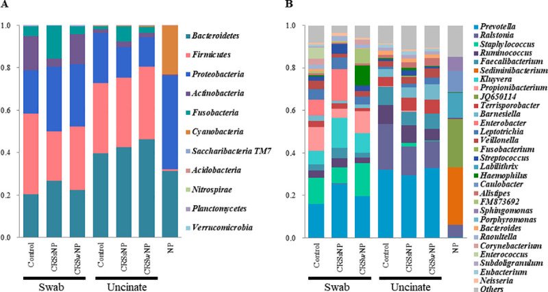
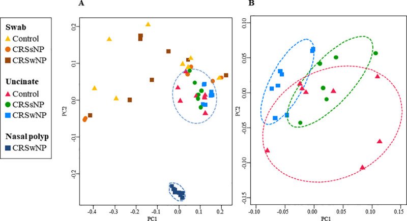
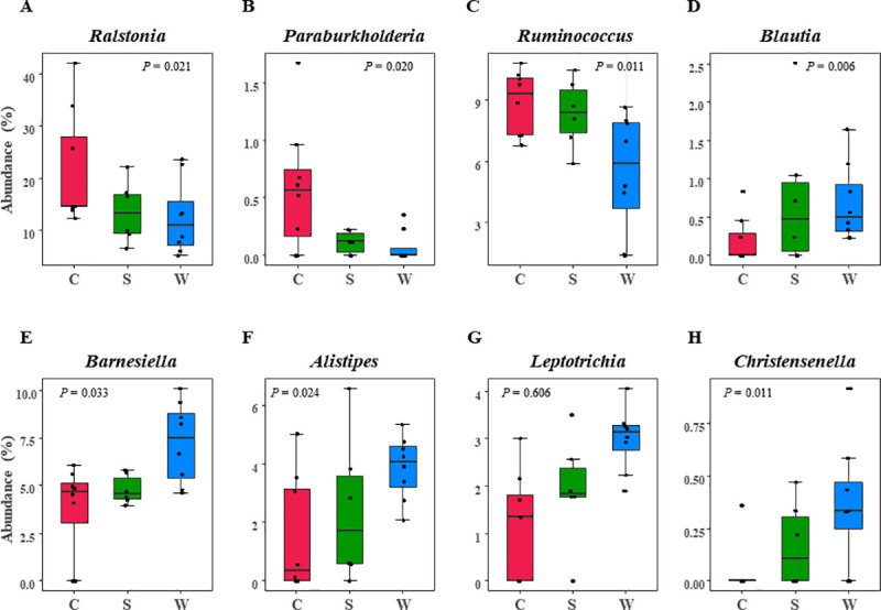
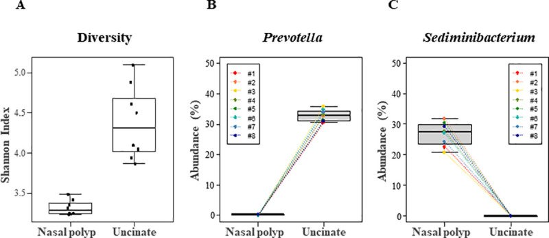
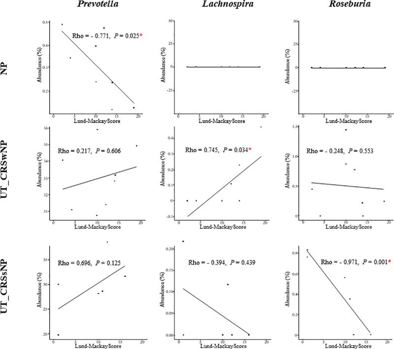
그림 출처: PubMed Central 공개 논문 (PMC8031401)
Conflict of interest statement
The authors have declared that no competing interests exist
그 밖의 정보
- 출판 형태
- Journal Article, Research Support, Non-U.S. Gov't
- 저널
- PloS one · eISSN 1932-6203
- 전자 출판
- 2021-04-08
- MeSH
- Adult · Bacteria / genetics · Biopsy / methods · Chronic Disease · Female · Humans · Male · Microbiota / genetics · Middle Aged · Nasal Polyps / microbiology · RNA, Ribosomal, 16S / genetics · Sinusitis / microbiology · Specimen Handling / methods
- Substances
- RNA, Ribosomal, 16S
- 참고문헌
- 43편