
Arthritis Res Ther. 2014;16(3):R126.
Therapeutic effect of anti-C-X-C motif chemokine 10 (CXCL10) antibody on C protein-induced myositis mouse.
제1저자 김진현 · 교신저자 송영욱
Abstract
INTRODUCTION C-X-C motif chemokine 10 (CXCL10) is a chemokine that plays a critical role in the infiltration of T cells in autoimmune diseases and is reported to be expressed in muscle tissue of polymyositis. To determine the therapeutic efficacy of CXCL10 blockade, we investigated the role of CXCL10 and the effect of anti-CXCL10 antibody treatment in C protein-induced myositis (CIM), an animal model of polymyositis.
METHODS CIM was induced with human skeletal muscle C protein fragment in female C57BL/6 mice. Immunohistochemistry of CXCL10 and C-X-C motif chemokine receptor 3 (CXCR3) and measurement of serum CXCL10 were performed. Cell surface markers and interferon-gamma (IFN-γ) and tumor necrosis factor-alpha (TNF-α) in CIM lymph node cells was investigated by flow cytometry. Mice with CIM were treated with anti-CXCL10 antibody or control antibody (anti-RVG1) and the inflammation in muscle tissue was assessed.
RESULTS Immunohistochemistry showed increased expression of CXCL10 and CXCR3 in the inflammatory lesions of muscle in CIM. Especially, CD8+ T cells invading myofiber expressed CXCR3. Serum level of CXCL10 was increased in CIM compared to the level in normal mice (normal mouse, 14.3 ± 5.3 pg/ml vs. CIM, 368.5 ± 135.6 pg/ml, P < 0.001). CXCR3 positivity in CD8+ T cells was increased compared to that of CD4+ T cells in the lymph node cells of CIM (CXCR3+ among CD8+ T cell, 65.9 ± 2.1% vs. CXCR3+ among CD4+ T cell, 23.5 ± 4.7%, P <0.001). Moreover, IFN-γ+ cells were increased among CXCR3+CD8+ T cells compared to CXCR3-CD8+ T cells (CXCR3+CD8+ T cell, 28.0 ± 4.2% vs. CXCR3-CD8+ T cell, 9.5 ± 1.5%, P = 0.016). Migration of lymph node cells was increased in response to CXCL10 (chemotactic index was 1.91 ± 0.45). CIM mice treated with anti-CXCL10 antibody showed a lower inflammation score in muscles than those with anti-RVG1 (median, anti-CXCL10 treatment group, 0.625 vs. anti-RVG1 treatment group, 1.25, P = 0.007).
CONCLUSIONS CXCL10/CXCR3 expression was increased in the inflammation of CIM model and its blockade suppressed inflammation in muscle.
Figures
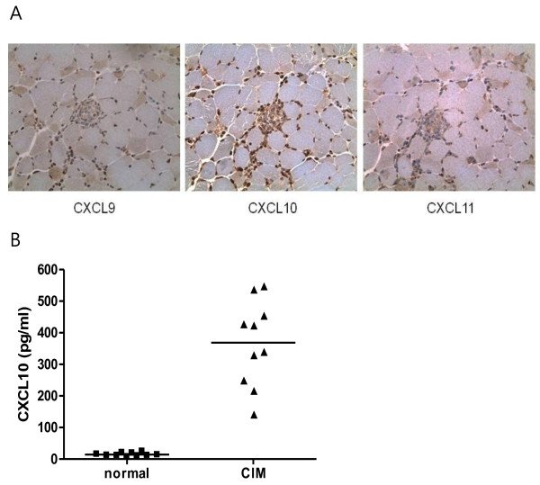
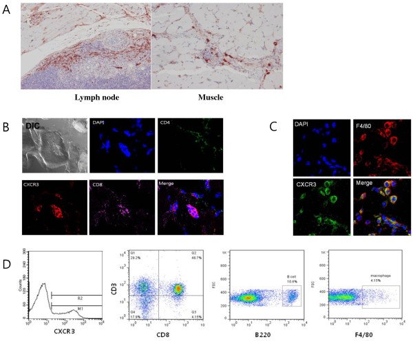
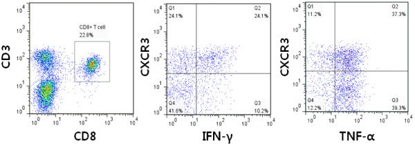
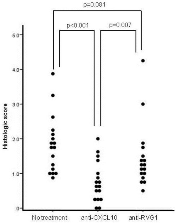
그림 출처: PubMed Central 공개 논문 (PMC4095607)
그 밖의 정보
- 출판 형태
- Journal Article, Research Support, Non-U.S. Gov't
- 저널
- Arthritis research & therapy · eISSN 1478-6362
- 전자 출판
- 2014-06-17
- MeSH
- Animals · Antibodies, Blocking / immunology / pharmacology · Carrier Proteins · Chemokine CXCL10 / antagonists & inhibitors / immunology / metabolism · Disease Models, Animal · Female · Flow Cytometry · Humans · Immunohistochemistry · Interferon-gamma / metabolism · Mice, Inbred C57BL · Myositis / blood / chemically induced / drug therapy · Polymyositis / blood / drug therapy · Receptors, CXCR3 / metabolism · Treatment Outcome · Tumor Necrosis Factor-alpha / metabolism · Myosin Binding Protein C
- Substances
- Antibodies, Blocking · Carrier Proteins · Chemokine CXCL10 · Interferon-gamma · Receptors, CXCR3 · Tumor Necrosis Factor-alpha · Myosin Binding Protein C · Cxcr3 protein, mouse
- 참고문헌
- 53편