
BMC Cancer. 2016;16:116.
Predictive and prognostic value of PET/CT imaging post-chemoradiotherapy and clinical decision-making consequences in locally advanced head & neck squamous cell carcinoma: a retrospective study.
제1저자 김률, 옥찬영 · 교신저자 김범석
소속 (17)
- Department of Internal Medicine, Seoul National University Hospital, 101 Daehak-ro, Jongno-gu, 110-744, Seoul, Korea. chrono0707@icloud.com.
- Department of Internal Medicine, Seoul National University Hospital, 101 Daehak-ro, Jongno-gu, 110-744, Seoul, Korea. ock.chanyoung@gmail.com.
- Department of Internal Medicine, Seoul National University Hospital, 101 Daehak-ro, Jongno-gu, 110-744, Seoul, Korea. bhumsuk@snu.ac.kr.
- Cancer Research Institute, Seoul National University College of Medicine, Seoul, Korea. bhumsuk@snu.ac.kr.
- Department of Internal Medicine, Seoul National University Hospital, 101 Daehak-ro, Jongno-gu, 110-744, Seoul, Korea. gabriel9@snu.ac.kr.
- Cancer Research Institute, Seoul National University College of Medicine, Seoul, Korea. gabriel9@snu.ac.kr.
- Department of Radiation Oncology, Seoul National University Hospital, Seoul, Korea. jinho.kim.md@gmail.com.
- Department of Nuclear Medicine, Seoul National University Hospital, Seoul, Korea. paengjc@snu.ac.kr.
- Department of Otorhinolaryngology, Seoul National University Hospital, Seoul, Korea. otolarynx@hanmail.net.
- Department of Otorhinolaryngology, Seoul National University Hospital, Seoul, Korea. jhunhah@gmail.com.
- Department of Otorhinolaryngology, Seoul National University Hospital, Seoul, Korea. kwontk@snu.ac.kr.
- Department of Internal Medicine, Seoul National University Hospital, 101 Daehak-ro, Jongno-gu, 110-744, Seoul, Korea. kimdw@snu.ac.kr.
- Cancer Research Institute, Seoul National University College of Medicine, Seoul, Korea. kimdw@snu.ac.kr.
- Department of Radiation Oncology, Seoul National University Hospital, Seoul, Korea. wuhg@snuh.org.
- Department of Otorhinolaryngology, Seoul National University Hospital, Seoul, Korea. mwsung@snu.ac.kr.
- Department of Internal Medicine, Seoul National University Hospital, 101 Daehak-ro, Jongno-gu, 110-744, Seoul, Korea. heo1013@snu.ac.kr.
- Cancer Research Institute, Seoul National University College of Medicine, Seoul, Korea. heo1013@snu.ac.kr.
Abstract
BACKGROUND The accuracy of (18)F-fluorodeoxygluocose positron emission tomography/computed tomography (PET/CT) in predicting immediate failure after radical chemoradiotherapy (CRT) for HNSCC is poorly characterized at present. The purpose of this study was to examine PET/CT as a predictive and prognostic gauge of immediate failure after CRT and determine the impact of these studies on clinical decision making in terms of salvage surgery.
METHODS Medical records of 78 consecutive patients receiving radical CRT for locally advanced HNSCC were reviewed, analyzing PET/CTs done before and 3 months after CRT. Immediate failure was defined as residual disease or locoregional and/or systemic relapse within 6 months after CRT.
RESULTS Maximum standard uptake value (SUV) of post CRT PET/CT (postSUVmax) was found optimal for predicting immediate failure at a cutpoint of 4.4. Sensitivity, specificity, negative predictive value (NPV), and positive predictive value (PPV) were 90.0%, 83.8%, 98.3%, and 45.0%, respectively. Of 78 patients studied, postSUVmax ≥ 4.4 prevailed in 20 (25.6%), with postSUVmax <4.4 in 58 (74.4%). At postSUVmax ≥ 4.4 (vs. postSUVmax <4.4) OS was poorer by comparison (3-year OS: 56.9 vs. 87.7%; P = 0.005), as was progression-free survival (3-year PFS: 42.9 vs. 81.1%; P < 0.001). At postSUVmax ≥ 4.4, OS with and without immediate salvage surgery did not differ significantly (3-year OS: 60.0 vs. 55.6%; Log-rank P = 0.913).
CONCLUSION Post CRT PET/CT imaging has prognostic value in terms of OS and PFS and is useful in predicting immediate therapeutic failure, given its high NPV. However, OS was not significantly altered by early salvage surgery done on the basis of post CRT PET/CT findings.
Figures
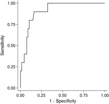
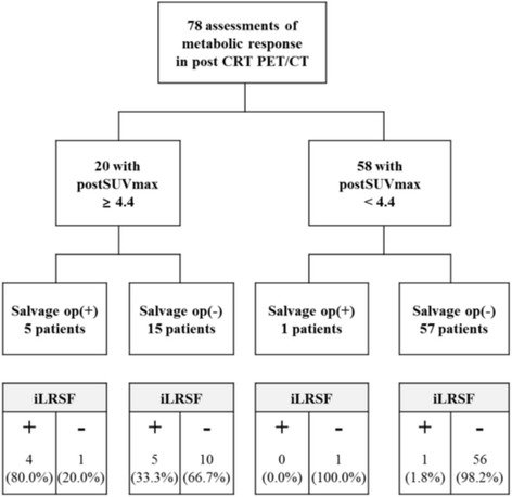
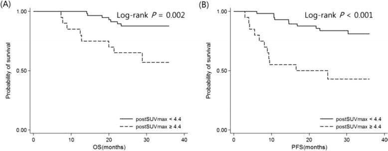
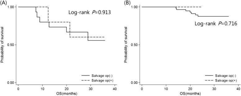
그림 출처: PubMed Central 공개 논문 (PMC4756525)
그 밖의 정보
- 출판 형태
- Journal Article, Research Support, Non-U.S. Gov't
- 저널
- BMC cancer · eISSN 1471-2407
- 전자 출판
- 2016-02-17
- MeSH
- Adult · Aged · Carcinoma, Squamous Cell / diagnosis / epidemiology / therapy · Female · Head and Neck Neoplasms / diagnosis / epidemiology / therapy · Humans · Image Interpretation, Computer-Assisted / methods · Male · Middle Aged · Positron-Emission Tomography / methods · ROC Curve · Retrospective Studies · Squamous Cell Carcinoma of Head and Neck · Survival Analysis · Tomography, X-Ray Computed / methods · Young Adult
- 참고문헌
- 35편