
Br J Cancer. 2008;98(6):1118-24.
High tumour islet macrophage infiltration correlates with improved patient survival but not with EGFR mutations, gene copy number or protein expression in resected non-small cell lung cancer.
제1저자 김동완 · 교신저자 허대석
소속 (1)
- Department of Internal Medicine, Seoul National University Hospital, Seoul, Korea.
Abstract
The purpose of this study was to investigate the prognostic value of tumour-associated macrophages with a focus on micro-anatomical localisation and determine whether molecular changes of the epidermal growth factor receptor (EGFR) are related to macrophage infiltration in resected non-small cell lung cancer (NSCLC). One hundred and forty-four patients were included in this study. Immunohistochemistry was used to identify CD68+ macrophages in the tumour islet and surrounding stroma. Epidermal growth factor receptor mutations were studied by direct sequencing. The EGFR gene copy number and protein expression were analysed by fluorescence in situ hybridisation and immunohistochemistry. Patients with a high tumour islet macrophage density survived longer than did the patient with a low tumour islet macrophage density (5-year overall survival rate was 63.9 vs 38.9%, P=0.0002). A multivariate Cox proportional hazard analysis revealed that the tumour islet macrophage count was an independent prognostic factor for survival (hazard ratio 0.471, 95% confidence interval 0.300-0.740). However, EGFR mutations, gene copy number, and protein expression were not related to the macrophage infiltration. In conclusion, tumour islet macrophage infiltration was identified as a strong favourable independent prognostic marker for survival but not correlated with the molecular changes of the EGFR in patients with resected NSCLC.
Figures
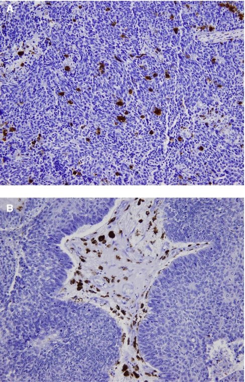
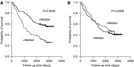
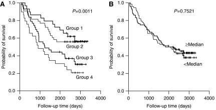
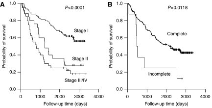
그림 출처: PubMed Central 공개 논문 (PMC2275476)
그 밖의 정보
- 출판 형태
- Journal Article, Research Support, Non-U.S. Gov't
- 저널
- British journal of cancer · ISSN 0007-0920
- 전자 출판
- 2008-02-19
- MeSH
- Aged · Antigens, CD / analysis · Antigens, Differentiation, Myelomonocytic / analysis · Carcinoma, Non-Small-Cell Lung / genetics / pathology · Cell Count · ErbB Receptors / metabolism · Female · Gene Dosage* · Genes, erbB-1* · Humans · Lung Neoplasms / genetics / pathology · Macrophages / immunology · Male · Middle Aged · Mutation* · Prognosis · Survival Analysis · CD68 Molecule
- Substances
- Antigens, CD · Antigens, Differentiation, Myelomonocytic · ErbB Receptors · CD68 antigen, human · CD68 Molecule
- 참고문헌
- 24편