National Research Laboratory for Stem Cell Niche, Center for Medical Innovation, Seoul National University Hospital, Seoul, Republic of Korea.
Center of Cell- and Bio-Therapy (CBT), Seoul National University Hospital, Seoul, Republic of Korea.
Department of Convergence Medicine, Pusan National University School of Medicine, Yangsan, Republic of Korea.
Department of Molecular Medicine and Biopharmaceutical Sciences, Graduate School of Convergence Science and Technology, and College of Medicine or College of Pharmacy, Seoul National University, Seoul, Republic of Korea.
Department of Pathology, Seoul National University College of Medicine, Seoul, Republic of Korea.
Department of Anatomy, Seoul National University College of Medicine, Seoul, Republic of Korea.
Genomics Core Facility, Department of Transdisciplinary Research and Collaboration, Biomedical Research Institute, Seoul National University Hospital, Seoul, Republic of Korea.
ExoCoBio Inc, Gasan digital 1-ro, Geumcheon-gu, Seoul, 08594, Republic of Korea.
Creative Research Initiative Center for Chromatin Dynamics, School of Biological Sciences, Seoul National University, Seoul, 151-742, Republic of Korea.
National Research Laboratory for Stem Cell Niche, Center for Medical Innovation, Seoul National University Hospital, Seoul, Republic of Korea. usahyosoo@gmail.com.
Center of Cell- and Bio-Therapy (CBT), Seoul National University Hospital, Seoul, Republic of Korea. usahyosoo@gmail.com.
Department of Molecular Medicine and Biopharmaceutical Sciences, Graduate School of Convergence Science and Technology, and College of Medicine or College of Pharmacy, Seoul National University, Seoul, Republic of Korea. usahyosoo@gmail.com.
Department of Internal Medicine, Seoul National University Hospital, 101 Daehak-ro, Jongno-gu, Seoul, 110-744, Korea. usahyosoo@gmail.com.
Abstract
BACKGROUND Little is known about endogenous inhibitors of angiogenic growth factors. In this study, we identified a novel endogenous anti-angiogenic factor expressed in pericytes and clarified its underlying mechanism and clinical significance.
METHODS Herein, we found Kai1 knockout mice showed significantly enhanced angiogenesis. Then, we investigated the anti-angiogenic roll of Kai1 in vitro and in vivo.
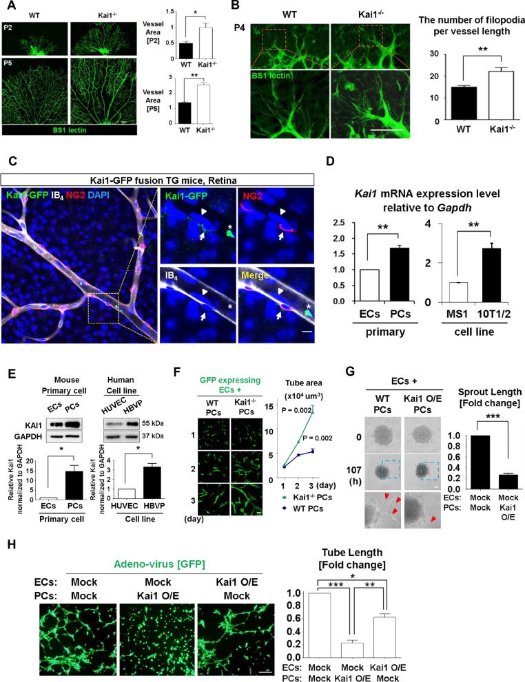
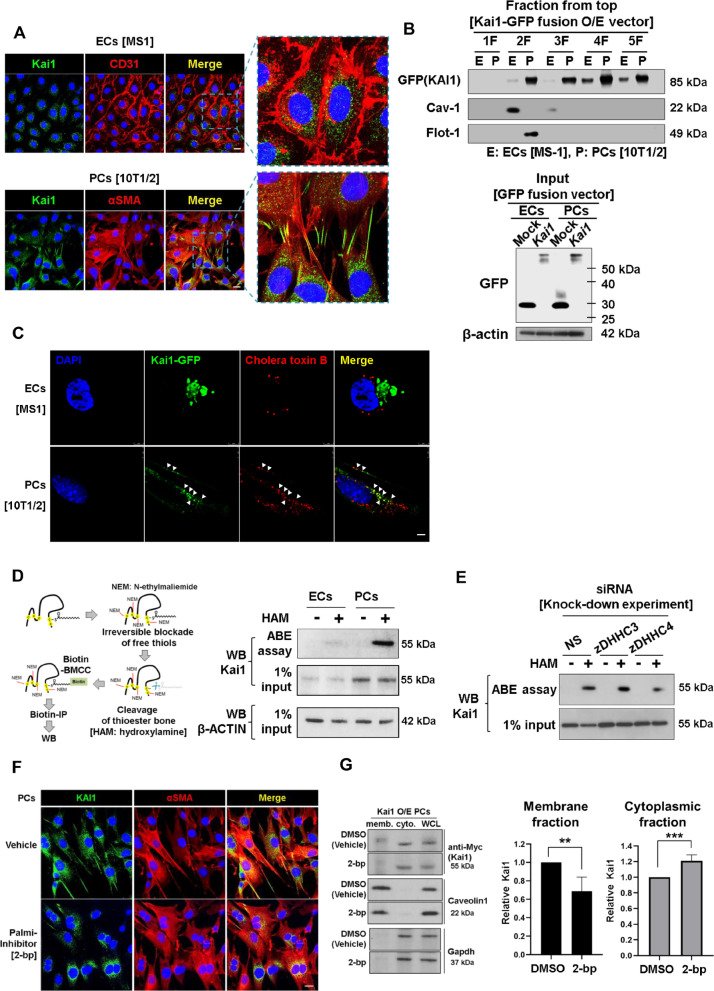
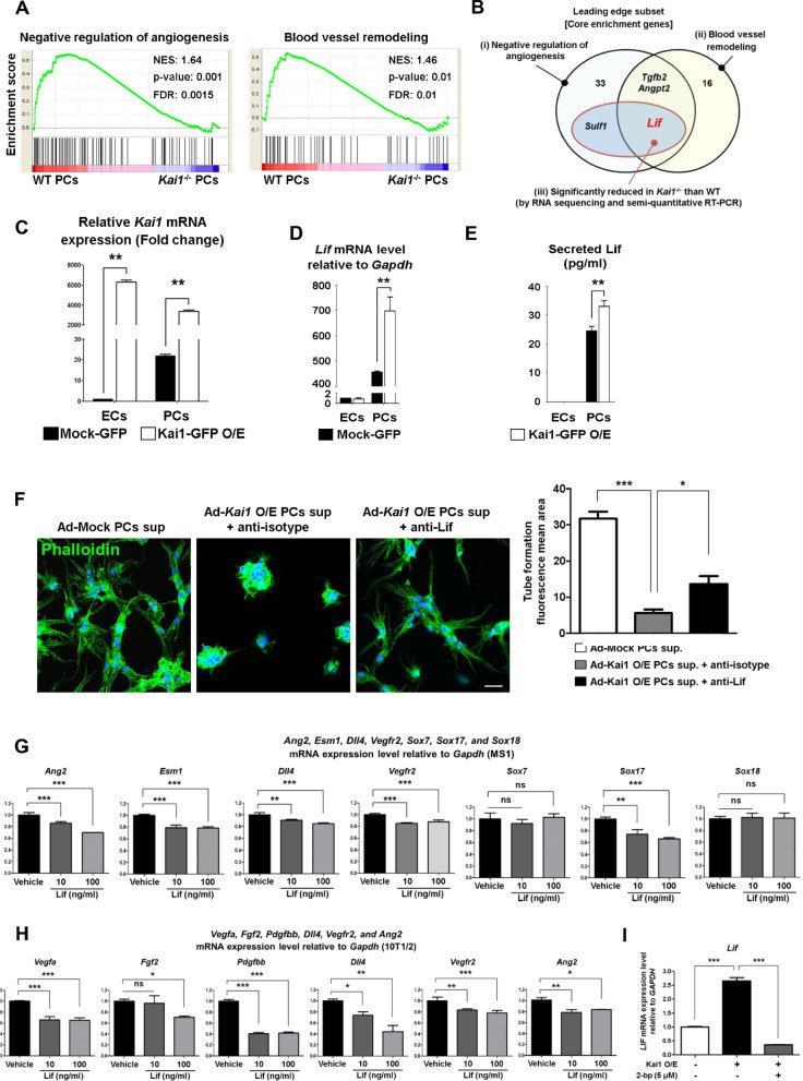
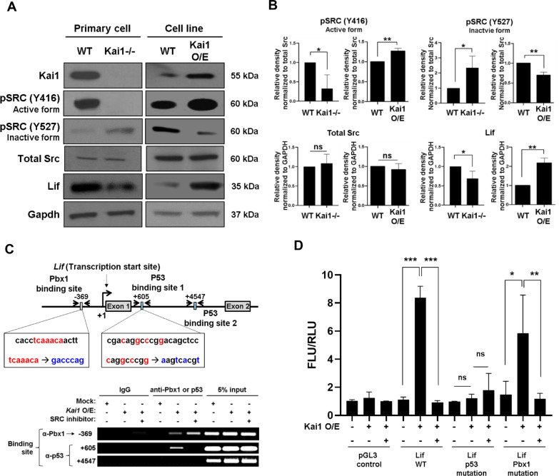
RESULTS KAI1 was mainly expressed in pericytes rather than in endothelial cells. It localized at the membrane surface after palmitoylation by zDHHC4 enzyme and induced LIF through the Src/p53 pathway. LIF released from pericytes in turn suppressed angiogenic factors in endothelial cells as well as in pericytes themselves, leading to inhibition of angiogenesis. Interestingly, KAI1 had another mechanism to inhibit angiogenesis: It directly bound to VEGF and PDGF and inhibited activation of their receptors. In the two different in vivo cancer models, KAI1 supplementation significantly inhibited tumor angiogenesis and growth. A peptide derived from the large extracellular loop of KAI1 has been shown to have anti-angiogenic effects to block the progression of breast cancer and retinal neovascularization in vivo.
CONCLUSIONS KAI1 from PC is a novel molecular regulator that counterbalances the effect of angiogenic factors.
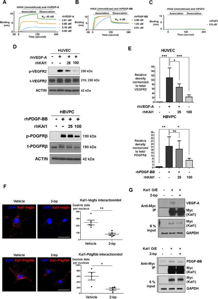
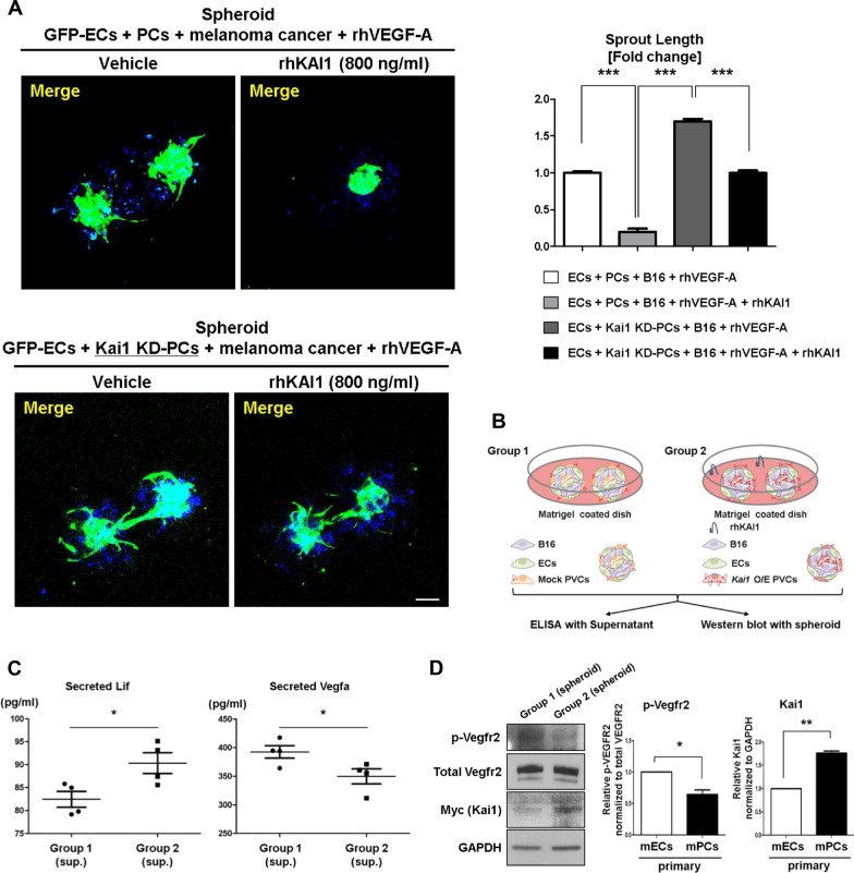
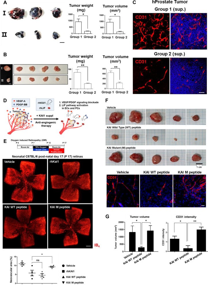
Fig. 1KAI1 was predominantly expressed in PCs rather than in ECs, and KAI1 expressed in PC has anti-angiogenic effects. a Left, retinal vasculature of the wild-type (WT) and Kai1 knockout (Kai1−/−) mice (postnatal day 2 and 5 days of age). Endothelial cells were stained with BS-I lectin. Scale bar, 100 μm. Right, quantification of the obtained results (Means ± SEM, 4–9 technical replicates). b Left, filopodia of retinal vessels in WT and Kai1−/− mice (postnatal day 4). Scale bar, 20 μm. Right, quantification of the obtained results (*p < 0.01, **p < 0.05, Means ± SEM, 3 or 4 technical replicates). c Tracking of Kai1 expressing cells in retina of the Kai1-GFP fusion knock-in mice. Confocal images of isolectin B4 (IB4, white), NG2 (red). Right, high-magnification images of the boxed area in the left panel Kai1 (green) was expressed in NG2 positive PCs (Arrow), but not ECs (Arrowhead). Asterisk indicates Kai1-expressing leukocyte or erythrocyte in vessel. Scale bar, 5 μm. dKai1 mRNA expression in mouse primary ECs and cell lines (MS1, endothelial cell line), and mouse primary PCs and cell line (10T1/2, pericyte cell line). **p < 0.05. e KAI1 protein expression in the mouse primary ECs, mouse primary PCs, human cell lines, HUVECs and HBVPs. *p < 0.01. f Left, in vitro co-culture matrigel tube formation assay using GFP expressing mouse ECs (MS1) co-cultured with Kai1−/− or WT mouse primary PCs. Scale bar, 100 μm. MS1 cells were transduced by lentivirus encoding GFP. Right, quantification of the obtained results (Means ± SEM, 7 technical replicates). g Left, In vitro matrigel tube formation assay, performed using co-culture spheroids, consisting of primary ECs and PCs, transduced with Myc-Kai1 vector and observed using time-lapse microscopy at the indicated time, 0 and 107 h. Scale bar, 50 μm. Right, quantification of the obtained results (***p < 0.001, Means ± SEM, 3 technical replicates). h Left, in vitro tube formation assay in MS1 and 10T1/2 co-cultured cells, transduced with the indicated adenovirus. Scale bar, 200 μm. Right, quantification of the obtained results (*p < 0.01, **p < 0.05, ***p < 0.001, Means ± SEM, 4 technical replicates)Fig. 2KAI1 localizes at the lipid rafts of PCs, which depends on palmitoylation. a Kai1 localization confirmed in MS1 and 10T1/2 cells using immunofluorescence. Scale bar, 50 μm. b Lipid rafts isolated from MS1 and 10T1/2 cells transfected with Kai1-GFP fusion vector using OptiPrep medium (frozen and crosslinks method). Flotillin-1 (planar type) and caveolin-1 (caveolae type) was detected in the lipid-raft membrane fraction. Fraction 1: no cellular protein. Fraction 2; Lipid raft proteins. Fraction 3; Non-raft membrane proteins. Fraction 4 and 5; Cytosolic proteins. Input samples are shown below. c Lipid raft marker (Cholera toxin B, red) co-localization with Kai1 (green) on MS1 and 10T1/2 cells transfected with Kai1-GFP fusion vector, observed using structured illumination microscopy. Arrowheads, co-localization of Kai1 protein and cholera toxin B. Scale bar, 2 μm. d Kai1 palmitoylation was analyzed by Acyl-Biotin Exchange (ABE) assay in ECs (MS1) and PCs (10T1/2). e zDHHC3 and zDHHC4 knockdown in 10T1/2, and their effects on Kai1 palmitoylation levels. f Changes in Kai1 localization after inhibition of palmitoylation using 2-bromopalmitate (2-bp) in 10T1/2. Scale bar, 50 μm. g Left, changes in Kai1 localization in the membrane and cytosol of 10T1/2 cells following the inhibition of palmitoylation using 2-bp. Right, quantification of the obtained results (**p < 0.05, ***p < 0.001, Means ± SEM, 3 technical replicates)Fig. 3Anti-angiogenic LIF is the downstream effect molecule induced by KAI1. a GSEA of the “negative angiogenic regulators” (GO: 0016525) and “blood vessel remodelling” (GO: 0001974), based on the RNA sequencing of WT and Kai1−/− PC. b Venn diagram showing the leading edge subset (core enrichment genes) of GSEA data for “negative angiogenic regulators” (GO: 0016525) and “blood vessel remodelling” (GO: 0001974), based on the RNA sequencing and semi-quantitative RT-PCR of WT and Kai1−/− PC. c qRT-PCR analysis for Kai1 mRNA level in Kai1-O/E ECs (MS1) and PCs (10T1/2). The genes were normalized by Gapdh (**p < 0.05, Means ± SEM, 3 technical replicates). d Lif expression in the Kai1-O/E MS1 and 10T1/2 cells (Means ± SEM, 3 technical replicates). e Lif level determination in the conditioned media of Kai1-O/E MS1 and 10T1/2 cells (Means ± SEM, 5 technical replicates). f Top, Tube formation assay using MS1 cells cultured in the conditioned media obtained from the mock and Kai1-O/E 10T1/2. These media were treated with control IgG or Lif antibody. Scale bar, 50 μm. Bottom, Quantification of the obtained results (*p < 0.01, **p < 0.05, ***p < 0.001, Means ± SEM, 3 technical replicates). g Changes in the levels of angiogenic molecules (Ang2, Esm1, Dll4, Vegfr2, Sox7, Sox17, and Sox18) by using semi-quantitative RT-PCR in MS1 cells following the treatment with mouse Lif (10 and 100 ng/mL). ns; not significant, **p < 0.05, ***p < 0.001. h Changes in Vegfa, Fgf2, Pdgfbb, Vegfr2, Dll4 and Ang2 levels in 10T1/2 cells in response to the Lif treatment (10 and 100 ng/mL). ns; not significant, *p < 0.01, **p < 0.05, ***p < 0.001. i After palmitoylation inhibition with 2-bp in 10T1/2 cells, Lif expression was detected by qRT-PCR. ***p < 0.001Fig. 4LIF is induced by KAI1 through Src/p53 axis in PCs. a Src phosphorylation levels and positions and Lif immunoblotting results in left, WT primary PC and Kai1−/− primary PC and, right, WT 10T1/2 and Kai1-O/E 10T1/2. b Quantification of the obtained results (ns not significant, *p < 0.01, **p < 0.05, Means ± SEM, 3 technical replicates). c Top, scheme, showing the regions targeted with the chromatin immune-precipitation (ChIP) assay and mutagenesis. Bottom, binding of p53 and Pbx1 on Lif gene-regulating element or promoter region using ChIP assay. d Activation of mouse Lif gene-regulating element or promoter in Kai1-O/E 10T1/2 cells using luciferase assay. ns not significant, *p < 0.01, **p < 0.05, ***p < 0.001Fig. 5KAI1 binds VEGF-A and PDGF-BB directly, resulting in inhibition of VEGFR2 and PDGFRβ signaling. a–c Protein–protein interactions between rhKAI1, rhVEGF-A, rhPDGF-BB and rhFGF2 observed using surface plasmon resonance. A shift in nm indicates the binding of two proteins. d Top, VEGFR2 phosphorylation levels in HUVECs, in response to rhVEGF-A and rhKAI1 treatment. Bottom, PDGFRβ phosphorylation level in human brain vascular PCs (HBVPs), in response to rhPDGF-BB and rhKAI1 treatment. e Quantification of the obtained results (ns not significant, *p < 0.01, **p < 0.05, ***p < 0.001, Means ± SEM, 3 technical replicates). f Left, Bimolecular fluorescent complementation assays using Duolink visualizes the interaction of Kai1-Vegfa and Kai1-Pdgfbb in living cells. Right, quantification of the obtained results (*p < 0.01, **p < 0.05, Means ± SEM, 3 technical replicates). Scale bar, 50 μm. g After palmitoylation inhibition in 293 T cells, protein–protein interactions between KAI1-rhVEGF-A (top) and KAI1-rhPDGF-BB (bottom) observed using the immunoprecipitation (IP) assayFig. 6Therapeutic potential of KAI1 to inhibit tumor angiogenesis and growth. a Left, In vitro matrigel tube formation assay using cellular spheroid consisting of MS1, 10T1/2 (WT and Kai1-KD), and B16 (melanoma cancer cell line) cells treated with 40 ng/mL of rhVEGF-A following the treatment with rhKAI1 (800 ng/mL). Scale bar, 100 μm. Right, Quantification of the obtained results (*p < 0.01, **p < 0.05, ***p < 0.001, Means ± SEM, 3 technical replicates). b Overview of experiment design applied to ELISA and western blot. c ELISA assay measuring the levels of mouse Lif and mouse Vegfa secreted into the media of three-hybrid spheres consisting of cancer (melanoma cells) and blood vessel cells, namely primary EC, as well as primary PC transfected with a mock vector (Group 1) and PC overexpressing Kai1 (Group 2). d Left, Vegfr2 phosphorylation levels and Kai1 immunoblotting results in group 1 and 2. Right, quantification of the obtained results (*p < 0.01, **p < 0.05, Means ± SEM, 3 technical replicates)Fig. 7Therapeutic potential of KAI1 and peptide to inhibit angiogenesis in tumors and prevent OIR-induced neovascularization. a B16 cells were subcutaneously injected in C57BL/6 mice with or without KAI1 supplement [combination of [1] Kai1-O/E 10T1/2 supernatant to provide anti-angiogenic LIF and [2] rhKAI1 to directly bind and inhibit VEGF and PDGF]. Left, tumor growth images are presented. Scale bar, 5 mm. Right, quantification of the obtained results (Means ± SEM, 3 biological replicates). b PC3 (human prostate cancer cell line) cells were subcutaneously injected into NSG mice in combination with or without KAI1 supplement. Left, tumor growth images are presented. Scale bar, 5 mm. Right, quantification of the obtained results (Means ± SEM, 5 biological replicates). c CD31 (red) immunostaining, and DAPI (blue) staining of human prostate (PC3) tumors treated with or without KAI1 supplement. Scale bar, 100 μm. d Schematic figure of dual anti-angiogenic effect of KAI1 in tumors. e Neonatal C57BL/6 mice with OIR at postnatal day 15 (P 15) were treated or not treated with a single intravitreal injection (1 μL) of vehicle only [PBS] (n = 4 retinas), rhKAI1 (600 ng), wild-type peptide KAI WT (200 ng) (n = 4 retinas), or mutant peptide KAI M (200 ng) (n = 4 retinas). Whole-mount retinas were stained with isolectin-B4 conjugated to Alexa Fluor 594 red-fluorescent dye. Top, Representative confocal microscopy images of retinas of OIR neonatal C57BL/6 mice at P17. Bottom, quantification of neovascularization in the retinas of OIR neonatal C57BL/6 mice treated or not treated with rhKAI1, KAI WT or KAI M peptides. Scale bar, 500 μm. f Mice were injected with matrigel and MDA-MB 231 cells in the presence of PBS alone (vehicle) or 200 ng KAI WT or M peptide (n = 5). After 23 days, the plugs were removed and the angiogenic responses were evaluated. Top, tumor volume was presented and bottom, representative IF images of tumors are shown. g Quantitative results of tumor volume (left) and CD31 intensity (right). Endothelial cells were visualized with CD31 (red) immunostaining, and DAPI (blue) staining of human breast cancer (MDA-MB231) treated with KAI WT or KAI M peptides. Scale bar, 20 μm. ns not significant, *p < 0.01, **p < 0.05, ***p < 0.001
그림 출처: PubMed Central 공개 논문 (PMC8444549)
Conflict of interest statement
The authors declare no competing interests.
그 밖의 정보
출판 형태
Journal Article, Research Support, Non-U.S. Gov't
저널
Journal of hematology & oncology · eISSN 1756-8722