SCI 발표논문

SCI 발표논문

← SCI 발표논문 목록

Circ Res. 2022;131(10):792-806.

Sox17 Deficiency Promotes Pulmonary Arterial Hypertension via HGF/c-Met Signaling.

, , , , , , , , , , , , , , , , ,

제1저자 박찬순 · 교신저자 박준빈, 김인준

소속 (9)
  1. Graduate School of Medical Science and Engineering (C.S.P., S.H.K., H.Y.Y., J.-H.K., E.L., H.P., J.M.Y., T.W.N., J.H., Y.S.J., I.K.).
  2. Division of Cardiology, Department of Internal Medicine (C.S.P., Y.S.C., H.K., S.-P.L., J.-B.P.).
  3. Department of Internal Medicine, Justus-Liebig University Giessen, Member of the German Center for Lung Research (DZL), Germany (R.T.S.).
  4. Center for Precision Medicine, Seoul National University Hospital and Seoul National University College of Medicine, Republic of Korea (H.K., S.-P.L.).
  5. Division of Cardiology, Department of Internal Medicine, Chungnam National University Hospital, Daejeon, Republic of Korea (J.-H.P.).
  6. Department of Ophthalmology, Dongguk University Ilsan Hospital, Goyang, South Korea (J.MY.).
  7. Center for Nanoparticle Research, Institute for Basic Science, Seoul, Republic of Korea (S.-P.L.).
  8. Department of Pharmacology, Pusan National University School of Medicine, Busan, Republic of Korea (S.S.B.).
  9. Biomedical Research Center, Korea Advanced Institute of Science and Technology, Daejeon, Republic of Korea (J.H., Y.S.J., I.K.).

Abstract

BACKGROUND In large-scale genomic studies, Sox17, an endothelial-specific transcription factor, has been suggested as a putative causal gene of pulmonary arterial hypertension (PAH); however, its role and molecular mechanisms remain to be elucidated. We investigated the functional impacts and acting mechanisms of impaired Sox17 (SRY-related HMG-box17) pathway in PAH and explored its potential as a therapeutic target.

METHODS In adult mice, Sox17 deletion in pulmonary endothelial cells (ECs) induced PAH under hypoxia with high penetrance and severity, but not under normoxia.

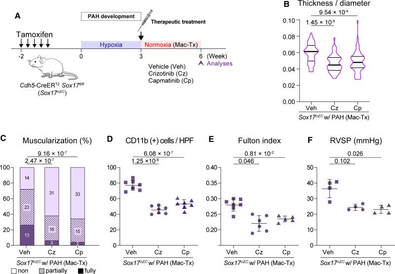
RESULTS Key features of PAH, such as hypermuscularization, EC hyperplasia, and inflammation in lung arterioles, right ventricular hypertrophy, and elevated pulmonary arterial pressure, persisted even after long rest in normoxia. Mechanistically, transcriptomic profiling predicted that the combination of Sox17 deficiency and hypoxia activated c-Met signaling in lung ECs. HGF (hepatocyte grow factor), a ligand of c-Met, was upregulated in Sox17-deficient lung ECs. Pharmacologic inhibition of HGF/c-Met signaling attenuated and reversed the features of PAH in both preventive and therapeutic settings. Similar to findings in animal models, Sox17 levels in lung ECs were repressed in 26.7% of PAH patients (4 of 15), while those were robust in all 14 non-PAH controls. HGF levels in pulmonary arterioles were increased in 86.7% of patients with PAH (13 of 15), but none of the controls showed that pattern.

CONCLUSIONS The downregulation of Sox17 levels in pulmonary arterioles increases the susceptibility to PAH, particularly when exposed to hypoxia. Our findings suggest the reactive upregulation of HGF/c-Met signaling as a novel druggable target for PAH treatment.

Keywords

RNAendothelial cellshypoxiapulmonary arterial hypertensionsequence analysis

Figures

Figure 1.
Figure 1. Endothelial Sox17 deficiency induces severe pulmonary arterial hypertension (PAH) with high penetrance in mice. A, Experimental schedule of Sox17 deficiency-induced PAH. Control and Sox17i∆EC mice were exposed to normoxia or hypoxia for 3 weeks. B–E, Sox17i∆EC mice exposed to hypoxia displayed typical pathologic features of PAH as shown by increased wall thickness (B) and hypermuscularization of distal pulmonary arterioles (n=50 per group, 5 arterioles in each mice; C), increased lung infiltration of CD11b+ cells (number per high power field of lung sections, n=7 for control/normoxic mice and n=8 for other groups; D), and RV hypertrophy measured by Fulton index (n=13 per group; E). F, Sox17i∆EC/hypoxic mice displayed hemodynamic evidence of PAH, including elevated RV systolic pressure, which was attenuated partially by overexpressing N1ICD in ECs (n=6 for control/normoxic and Sox17i∆EC/hypoxic mice, n = 5 for Sox17i∆EC/N1ICDiOE/hypoxic mice, and n=4 for other groups), and (G) D-shaped left ventricle (LV), which contrasted to normal shape of the LV in control mice. Asterisks and yellow lines indicate left ventricular cavity and endocardial border, respectively. Data are presented as mean±SD. Generalized estimating equation models (B and C), 1-way ANOVA with Scheffé post-test (D and E), and Kruskal-Wallis test with Dunn’s post-test (F) were performed. Multiple comparison was performed between Sox17i∆EC/hypoxic mice and each other group (F). Scale bar, 2 mm (G). Ctrl indicates control; HPF, high-power field; LV, left ventricle; and RVSP, RV systolic pressure.
Figure 2.
Figure 2. Validation of transcriptomic profiling of pulmonary endothelial cells (ECs). A, Experimental strategy for isolating endothelial-enriched transcripts in the lung. Cdh5-CreERT2; RPL22tm1.1Psam mice were maintained under normoxia as control group and Cdh5-CreER T2; Sox17fl/fl RPL22tm1.1Psam mice were exposed to hypoxia as the Sox17 i∆EC/hypoxic group following tamoxifen administration, which allowed the synthesis of hemagglutinin (HA)-tagged ribosomes specifically in ECs. Lungs were then harvested. B, Volcano plot of differentially expressed genes in lung ECs of control and Sox17i∆EC/hypoxic mice. Sox17 highlighted in blue confirms its decreased level. C, Immunofluorescence image verifying EC-specific HA immunostaining in the lung. D, Fold changes in transcripts of marker genes for EC and other cell types induced by capturing HA-tagged ribosomes. Transcript levels in lung tissue samples for RNA sequencing are validated by quantitative PCR (n=3). Relative levels of transcripts are normalized to those of Gapdh. As controls, the averages of samples before the precipitation of HA-tagged ribosomes (left column of each pair) are arbitrarily set as 1. EC markers (Pecam1, Cdh5) are enriched, whereas markers of other cell types (Pdgfrb, pericytes; Acta2, VSMCs; Sftpc, pneumocytes) are excluded. Data are presented as mean±SD. E, Three-dimensional principal component (PC) analysis and (F) unsupervised clustering clearly separate RNA sequencing data from the control (n=4) and Sox17i∆EC/hypoxic groups (n=4). Scale bar, 10 μm (C).
Figure 3.
Figure 3. Loss of Sox17 promotes abnormal proliferation and inflammatory activation in lung endothelial cells (ECs) under hypoxic stress. A, The top 5 enriched gene sets by gene ontology (GO) term analysis (top, biological process; bottom, reactome pathways). Gene sets associated with cell proliferation are displayed in dark orange and those associated with inflammation are displayed in light orange. B, Dot plot of GSEA results related to cell proliferation and inflammation. P are represented by color gradient and the false discovery rate adjusted P (ie, q-values) are depicted by circle size. C, Immunostaining for Ki67, a marker of proliferation, in distal pulmonary arterioles and its quantification in ECs (n=6 mice per group, 5 arterioles from each mouse). Note the significant increase in Ki67 expression in Sox17i∆EC/hypoxic mice versus control/normoxic mice. D, Histologic image showing vascular obliteration in pulmonary arterioles by abnormal stacking of ECs in Sox17i∆EC/hypoxic mice. E, Immunostaining for S100A4 in distal pulmonary arterioles and its quantification in ECs (n=5 mice per group, 5 arterioles from each mouse). Note the lack of S100A4 signals in ECs in both control/normoxic and Sox17i∆EC/hypoxic mice and no between-group difference. F, No significant transcriptional changes in mesenchymal markers on the basis of RNA-sequencing data. Data are presented as mean±SD. Numbers in parentheses refer to the numerator and denominator from which the relevant percentages are calculated (C and E). Generalized estimating equation models were adopted for clustered data (C and E). Scale bars: 10 μm (C, E), 20 μm (D).
Figure 4.
Figure 4. Inhibition of HGF (hepatocyte growth factor)/c-Met signaling prevents the development of pathologic changes promoted by Sox17 deficiency. A, The top-ranked reactome pathways by gene set enrichment analysis (GSEA; left) and GSEA plot showing negative enrichment of negative regulation (ie, activation) of c-Met signaling (right) in Sox17i∆EC/hypoxic mice. B, Representative images of HGF upregulation in distal pulmonary arterioles of Sox17i∆EC/hypoxic mice. C, The percentage of HGF+ endothelial cells (ECs) in distal pulmonary arteriolar ECs in each experimental mouse group (n=6 mice per group, 5 arterioles from each mouse). D, Experimental schedule for c-Met inhibition in a preventive setting: following tamoxifen injection, Sox17i∆EC mice treated with vehicle (Veh, 10% DMSO), macitentan (Mac, a vasodilator), crizotinib (Cz, a c-Met inhibitor), or a combination of macitentan and crizotinib (Mac/Cz) during hypoxic exposure for 3 weeks and control mice treated with vehicle under normoxia for the same period. E–H, Attenuation of PAH phenotypes by c-Met inhibition combined with macitentan comparable to control mice as shown by reduced wall thickness (E) and muscularization of distal pulmonary arterioles (n=100 per group, 5 arterioles in each mice; F), attenuated lung infiltration by CD11b+ cells (number per high power field, n=6 for control/normoxic mice and n=7 for other groups; G), and improved RV hypertrophy (n=9 for control/normoxic and Sox17i∆EC/hypoxic groups, n=8 for Sox17i∆EC/hypoxic mice treated with macitentan and Sox17i∆EC/hypoxic mice treated with macitentan and crizotinib, and n=7 for Sox17i∆EC/hypoxic mice treated with crizotinib; H) in Sox17i∆EC/hypoxic mice. Data are presented as mean±SD. Numbers in parentheses refer to the numerator and denominator from which the relevant percentages are calculated (C). Generalized estimating equation models (C, E, and F), 1-way ANOVA with Scheffé post-test (H), and Kruskal-Wallis test with Dunn’s post-test (G) were performed. Scale bars in B: 10 μm. ES indicates enrichment score; and HPF, high-power field.
Figure 5.
Figure 5. Therapeutic treatment with c-Met inhibitors reverses established pulmonary arterial hypertension (PAH). A, Experimental schedule for c-Met inhibition in a therapeutic setting: following tamoxifen injection, Sox17i∆EC mice were confirmed to have PAH by echocardiography imaging at the end of 3 weeks of hypoxia and then treated with vehicle (Veh, 10% DMSO), crizotinib (Cz, a c-Met inhibitor), or capmatinib (Cp, a c-Met inhibitor) in the presence of macitentan (Mac-Tx) during the subsequent 3 weeks of normoxia. B–F, Reversal of PAH phenotypes developed in Sox17i∆EC mice by c-Met inhibition combined with macitentan as shown by reduced wall thickness (B) and muscularization of distal pulmonary arterioles (n=50 per group, 5 arterioles in each mice; C), decreased lung infiltration by CD11b+ cells (number per high-power field, n=7 per group; D), improved RV hypertrophy (n=7 mice treated with macitentan, n=6 mice treated with macitentan and crizotinib, and n=5 mice treated with macitentan and capmatinib; E), and reduced RV systolic pressure (n=4 per group; F). Data are presented as mean±SD. Generalized estimating equation models (B and C), 1-way ANOVA with Scheffé post-test (D), and Kruskal-Wallis test with Dunn’s post-test (E and F) were performed. HPF indicates high-power field; and PAH, pulmonary arterial hypertension.
Figure 6.
Figure 6. Repressed Sox17 expression and increased HGF (hepatocyte grow factor) expression in pulmonary arterial endothelial cells (ECs) from patients with pulmonary arterial hypertension (PAH) in comparison with control subjects. A, Immunostaining for Sox17 in lung tissue sections from a control subject and a PAH patient. Note the repressed Sox17 expression in arteriolar ECs (yellow arrow) and hypermuscularization (white arrowhead) in the PAH patient. B, Proportions of Sox17 expression levels in pulmonary arteriolar ECs. Note the substantial proportion of low Sox17 expression in the PAH group, contrasting to the control group in which no case of low Sox17 expression was found. C, Immunostaining of HGF in lung tissue sections from a control subject and a patient with PAH. Note the increased HGF expression in arteriolar ECs (yellow arrow) and hypermuscularization (white arrowhead) in the patient with PAH. D, Proportions of HGF expression levels in pulmonary arteriolar ECs. Note the substantial proportion of high HGF expression in the PAH group, contrasting to the control group in which no case of high HGF expression was found. Numbers in bars indicate absolute numbers of cases. Demographic data for each subject are presented in Table S1. Scale bars in A and C: 10 μm.
Figure 7.
Figure 7. Summarizing illustration. Schematic diagram depicting the role of Sox17 in pulmonary arterial health and its loss leading to pulmonary arterial hypertension via HGF (hepatocyte grow factor) upregulation and impaired Notch signaling. EC indicates endothelial cell; and PAH, pulmonary arterial hypertension.

그림 출처: PubMed Central 공개 논문 (PMC9612711)

Comment · 정정

  • Comment in 789
  • Comment in 790
그 밖의 정보
출판 형태
Journal Article, Research Support, Non-U.S. Gov't
저널
Circulation research · eISSN 1524-4571
전자 출판
2022-10-07
MeSH
Animals · Mice · Endothelial Cells / metabolism · HMGB Proteins / metabolism · Hypertension, Pulmonary* / genetics / metabolism · Hypoxia / complications / metabolism · Pulmonary Arterial Hypertension* / genetics · Pulmonary Artery / metabolism · Signal Transduction · SOXF Transcription Factors / genetics / metabolism · Proto-Oncogene Proteins c-met / metabolism · Hepatocyte Growth Factor
Substances
HMGB Proteins · SOXF Transcription Factors · Proto-Oncogene Proteins c-met · Sox17 protein, mouse · HGF protein, mouse · Hepatocyte Growth Factor
참고문헌
55편