
Gut Liver. 2019;13(4):415-420.
A Newly Designed 3-Dimensional Printer-Based Gastric Hemostasis Simulator with Two Modules for Endoscopic Trainees (with Video).
제1저자 이동석 · 교신저자 이진혁, 안지용
소속 (2)
- Department of Gastroenterology, SMG-SNU Boramae Medical Center, Seoul National University of College of Medicine, Seoul, Korea.
- Department of Gastroenterology, Asan Medical Center, University of Ulsan College of Medicine, Seoul, Korea.
Abstract
BACKGROUND/AIMS We used 3-dimensional (3D) printing technology to create a new hemostasis simulator for the stomach and investigated its efficacy and realism in endoscopic hemostasis training.
METHODS A new stomach hemostasis simulator, with two hemostasis modules for hemoclipping and injection, was constructed using a 3D printer. Twenty-one endoscopists, including 11 first-year fellows (beginner group) and 10 faculty members (expert group), tested the performance of the simulator. We recorded and reviewed five training sessions and evaluated the simulator with questionnaires using a 7-point Likert scale.
RESULTS The mean evaluation score of the expert group was 6.3±0.5 for the hemoclipping module and 6.0±0.6 for the injection module. The expert group strongly agreed that endoscopic handling in the simulator was realistic and reasonable for hemostasis training. The mean procedure time for hemoclipping was 72.7±7.1 seconds for the beginner group and 19.7±1.2 seconds for the expert group. The mean procedure time for injection was 92.1±9.8 seconds for the beginner group and 36.3±2 seconds for the expert group. The procedure time of beginner group became shorter with repetition and was significantly lower by the fifth trial.
CONCLUSIONS A new 3D-printed hemostasis simulator is capable of hemostasis training and can very effectively train beginners before they perform the procedure in patients with gastrointestinal bleeding.
Keywords
EducationEndoscopyHemostasisPrintingSimulatorthree-dimensional
Figures
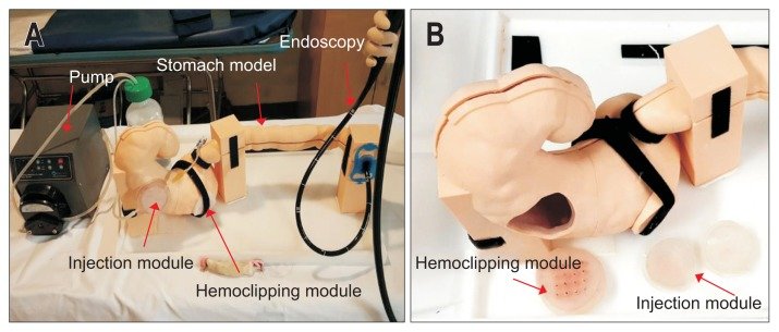
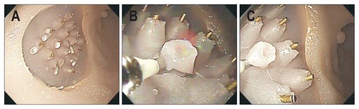
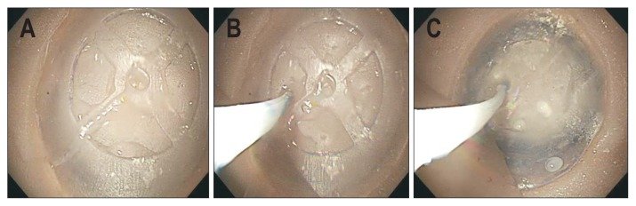
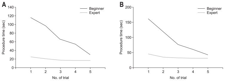
그림 출처: PubMed Central 공개 논문 (PMC6622563)
Conflict of interest statement
CONFLICTS OF INTEREST. No potential conflict of interest relevant to this article was reported.
그 밖의 정보
- 출판 형태
- Journal Article, Video-Audio Media
- 저널
- Gut and liver · eISSN 2005-1212
- MeSH
- Electrocoagulation · Gastroenterology / education · Gastrointestinal Hemorrhage / therapy · Gastroscopy / education · Hemostasis, Endoscopic / education · Humans · Models, Anatomic · Printing, Three-Dimensional* · Simulation Training* · Stomach
- 참고문헌
- 19편