
PLoS One. 2018;13(1):e0190261.
Liver volume-based prediction model stratifies risks for hepatocellular carcinoma in chronic hepatitis B patients on surveillance.
제1저자 이충섭 · 교신저자 김진욱
소속 (5)
- Department of Medicine, Seoul National University Bundang Hospital, Seongnam, Republic of Korea.
- Department of Internal Medicine, Seoul National University College of Medicine, Seoul, Republic of Korea.
- Department of Internal Medicine, SMG-SNU Boramae Medical Center, Seoul, Republic of Korea.
- Department of Gastroenterology, Ajou University School of Medicine, Suwon, Republic of Korea.
- Department of Radiology, Seoul National University College of Medicine, Seoul, Republic of Korea.
Abstract
BACKGROUND AND AIM The aim of this study was to determine whether dynamic computed tomography (CT)-measured liver volume predicts the risk of hepatocellular carcinoma (HCC) when the CT scans do not reveal evidence of HCC in chronic hepatitis B (CHB) patients on surveillance.
METHODS This retrospective multicentre cohort study included 1,246 patients who received entecavir and regular HCC surveillance in three tertiary referral centres in South Korea. Liver volumes were measured on portal venous phase CT images. A nomogram was developed based on Cox independent predictors and externally validated. Time-dependent receiver operating characteristic (ROC) analysis was performed for comparison with previous prediction models.
RESULTS Patients who received dynamic CT studies during surveillance had significantly higher risk for HCC compared to patients without CT studies (hazard ratio [HR] = 3.1; p < 0.001). Expected/measured liver volume ratio was an independent predictor of HCC (HR = 4.2; p = 0.002) in addition to age, sex and cirrhosis. The nomogram based on the four predictors discriminated risks for HCC (HR = 4.1 and 6.0 in derivation and validation cohort, respectively, for volume score > 150; p < 0.001). Time-dependent ROC analysis confirmed better performance of the volume score compared to HCC prediction models with conventional predictors (integrated area under curve = 0.758 vs. 0.661-0.712; p < 0.05).
CONCLUSIONS CT-measured liver volume is an independent predictor of future HCC, and nomogram-based liver volume score may stratify the risks of HCC in CHB patients who showed negative CT findings for HCC during surveillance.
Figures

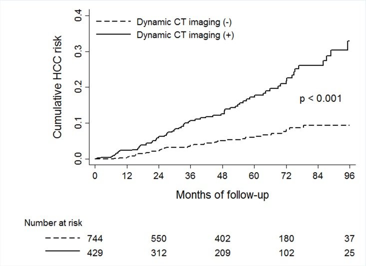
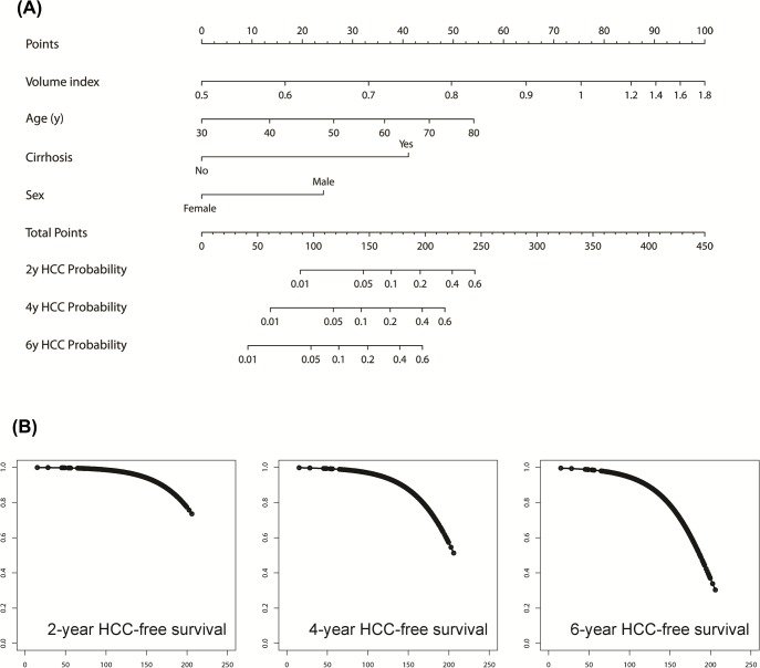
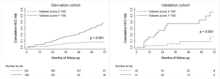
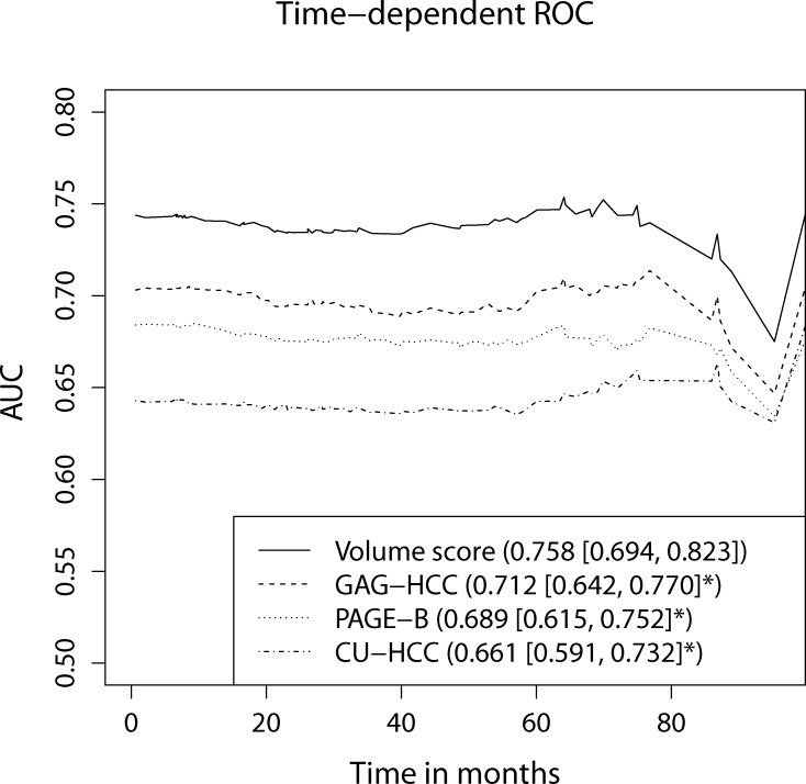
그림 출처: PubMed Central 공개 논문 (PMC5749771)
Conflict of interest statement
Competing Interests:The authors have declared that no competing interests exist.
그 밖의 정보
- 출판 형태
- Journal Article, Multicenter Study, Research Support, Non-U.S. Gov't
- 저널
- PloS one · eISSN 1932-6203
- 전자 출판
- 2018-01-02
- MeSH
- Adult · Aged · Carcinoma, Hepatocellular / complications / pathology · Female · Hepatitis B, Chronic / complications / pathology · Humans · Liver / diagnostic imaging / pathology · Liver Neoplasms / complications / pathology · Male · Middle Aged · Models, Biological · Risk Factors · Tomography, X-Ray Computed
- 참고문헌
- 36편