Biomedical Research Institute and IRICT, Seoul National University Hospital, 101 DaeHak-ro, JongRo-gu Seoul, 110-744, Republic of Korea.
Department of Internal Medicine and IRICT, Seoul National University Hospital, 101 DaeHak-ro, JongRo-gu Seoul, 110-744, Republic of Korea.
Department of Molecular Medicine and Biopharmaceutical Sciences, Graduate School of Convergence Science and Technology, Seoul National University, Korea.
Abstract
Skeletal muscle regeneration occurs continuously to repair muscle damage incurred during normal activity and in chronic disease or injury. Herein, we report that A-kinase anchoring protein 6 (AKAP6) is important for skeletal myoblast differentiation and muscle regeneration. Compared with unstimulated skeletal myoblasts that underwent proliferation, differentiated cells show significant stimulation of AKAP6 expression. AKAP6 knockdown with siRNA effectively halts the formation of myotubes and decreases the expression of the differentiation markers myogenin and myosin heavy chain. When shAKAP6-lentivirus is delivered to mice with cardiotoxin (CTX)-induced muscle injury, muscle regeneration is impaired compared with that of mice injected with control shMock-lentivirus. The motor functions of mice infected with shAKAP6-lentivirus (CTX+shAK6) are significantly worse than those of mice infected with shMock-lentivirus (CTX+shMock). Mechanistic analysis showed that AKAP6 promotes myogenin expression through myocyte enhancer factor 2A (MEF2A). Notably, myogenin increases AKAP6 expression as well. The results of chromatin immunoprecipitation and luciferase assays showed that myogenin binds to an E-box site on the AKAP6 promoter. Taken together, our findings demonstrate a novel interplay between AKAP6 and myogenin, and we suggest that AKAP6 is an important regulator of myoblast differentiation, myotube formation, and muscle regeneration.
Figures
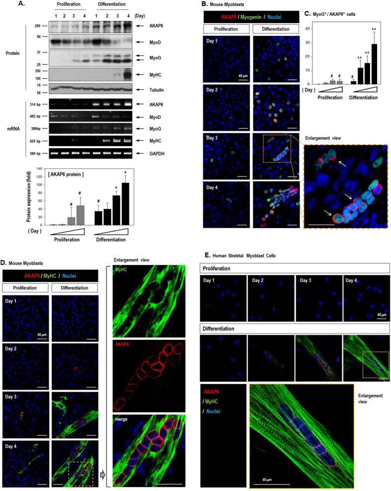
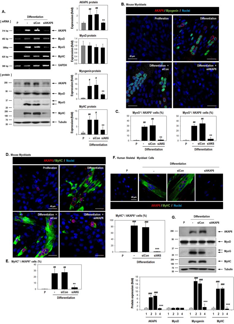
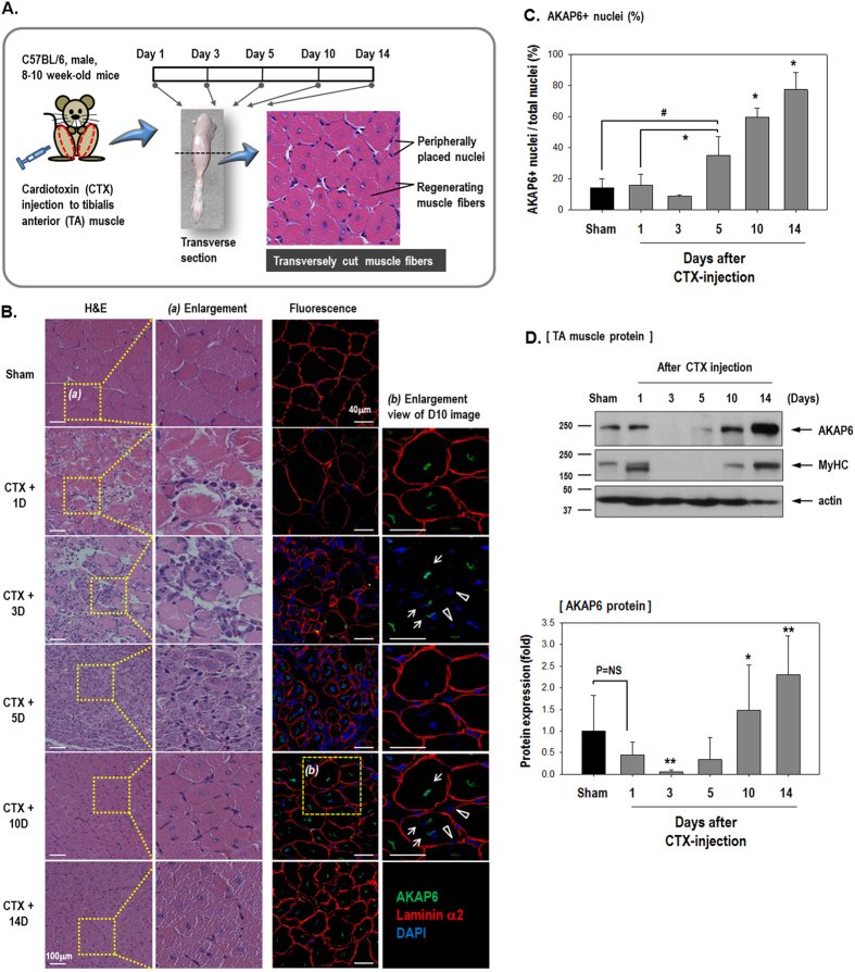
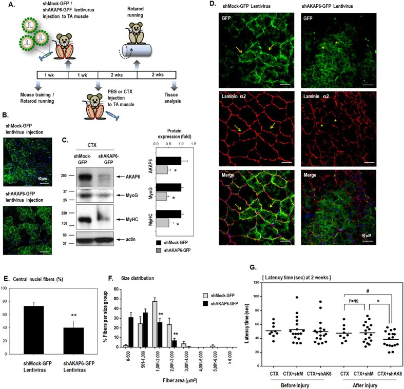
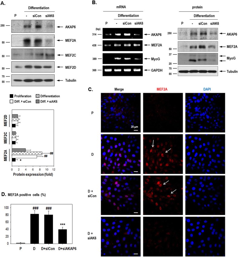
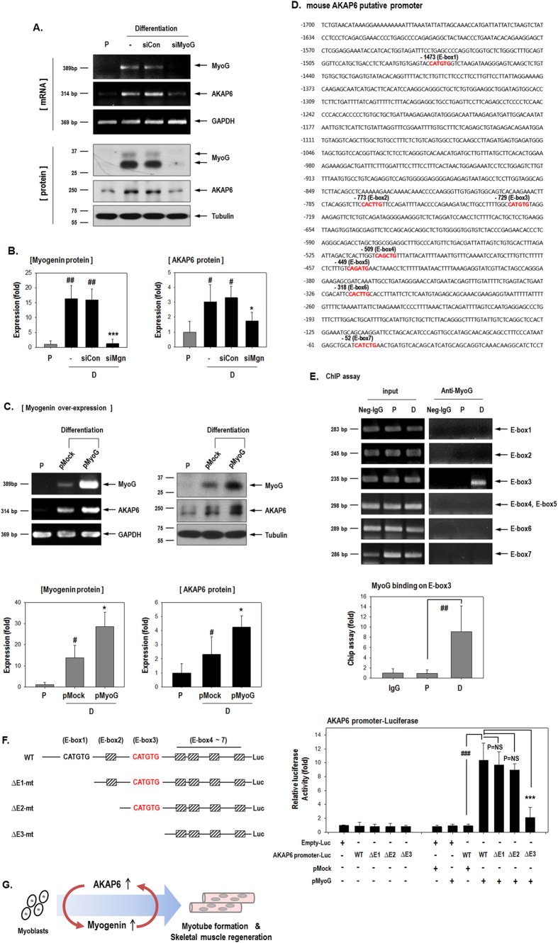
Figure 1 Expression pattern of AKAP6 and several differentiation markers in differentiating myoblasts. (A) Daily harvested protein and RNA samples from mouse C2C12 myoblasts analyzed by western blotting (Top) and RT-PCR (Middle). AKAP6 and the differentiation markers myogenin and MyHC increased during differentiation. The quantification graph shows the relative fold change compared with levels on day 1 under proliferative conditions (Bottom, n = 3). #p < 0.05 versus proliferation on day 1; *p < 0.05 versus differentiation on day 1 (one-way ANOVA). (B) C2C12 cells were stained with AKAP6 (Red) and myogenin (Green). AKAP6 staining is observed in the nuclear envelope (arrows), and myogenin is detected in the nucleus (Magnification: ×400). (C) Quantification graph for AKAP6+/Myogenin+ cells (n = 5) under proliferation (gray) or differentiation (black) conditions. #p < 0.05 versus proliferation on day 1; **p < 0.01 versus differentiation on day 1 (one-way ANOVA). (D) AKAP6-positive (Red) and MyHC-positive (Green) cells are mature myotubes (Magnification: ×400). (E) Immunofluorescence staining for AKAP6 (Red) and MyHC (Green) in human skeletal muscle myoblasts (HSMMs; magnification: ×400).Figure 2 AKAP6 depletion decreases myogenin and MyHC levels and impairs myotube formation. (A) Inhibition of AKAP6 expression by using an AKAP6 siRNA was confirmed with RT-PCR and western blotting. C2C12 cells were transfected with a specific siRNA against AKAP6 and further incubated under differentiation conditions for 3 days. Quantification graphs for western blotting are shown. The expression was normalized to that of the proliferation group and shown as fold changes (n = 3 each, right). #p < 0.05 and ##p < 0.01 versus proliferation; **p < 0.01 versus siCon + differentiation (one-way ANOVA). P, proliferation conditions; siCon, control siRNA; siAKAP6, siRNA against AKAP6. (B) C2C12 cells were transfected with siCon or siAKAP6, incubated in differentiation medium for 3 days, fixed, and immunofluorescence-stained for AKAP6 (Red) and myogenin (Green). Original magnification: ×400. (C) The graph shows the percentage of myogenin+/AKAP6+ cells or myogenin+/AKAP6− cells divided by the number of nuclei (n = 5 each). ##p < 0.01 versus proliferation; **p < 0.01 versus siCon + differentiation (one-way ANOVA). (D) Immunofluorescence staining for AKAP6 (Red) and MyHC (Green) after siAKAP6 transfection in C2C12 cells. Magnification: ×400. (E) The graph shows the percentage for MyHC+/AKAP+ cells divided by the number of nuclei (n = 5 each). ##p < 0.01 versus proliferation; **p < 0.01 versus siCon + differentiation (one-way ANOVA). (F) Effect of AKAP6 knockdown in human myoblasts, HSMMs, under differentiation conditions. After transfection with siAKAP6, HSMMs were stained for AKAP6 (Red) and MyHC (Green). Magnification: ×400. The graph shows the percentage for MyHC+/AKAP+ cells divided by the number of nuclei (n = 5 each). ###p < 0.001 versus proliferation; ***p < 0.01 versus siCon + differentiation (one-way ANOVA). (G) Western blotting for AKAP6 and myogenic genes in HSMMs. The expression was normalized to that of the proliferation group and is shown as fold change (n = 5 each, ###p < 0.001 versus proliferation, ***p < 0.001 versus differentiation + siCon). 1, Proliferation conditions; 2, Differentiation conditions; 3, Diff + siCon; 4, Diff + siAKAP6.Figure 3 AKAP6 expression during the muscle repair period after injury. (A) Timeline for the in vivo experiment. (B) Hematoxylin and eosin (H&E) or immunofluorescence staining of transverse muscle sections after cardiotoxin (CTX) injection to the tibialis anterior (TA) muscle on the indicated days. Sham sections were from PBS-injected muscles. H&E staining (Magnification: ×200). Immunofluorescence staining shows that AKAP6 (Green) is predominantly observed in a centrally located nuclear envelope (arrows). Nuclei in a peripheral position in mature myofibers did not express AKAP6 (arrowheads). Laminin α2 for muscle fiber (Red), DAPI for nuclei (Blue; magnification: ×400). Immunofluorescence staining was performed in four mice using four different sections per mouse. (C) The graph shows the percentage of AKAP6-expressing nuclei divided by the number of nuclei (n = 5 each). #p < 0.05 versus Sham; *p < 0.05 versus Day1 CTX (one-way ANOVA). (D) At the indicated time points, the TA muscles were frozen directly in liquid nitrogen, lysed in RIPA buffer, and analyzed with western blotting (Top). Quantification graph of the AKAP6 western blot shows fold changes from values in sham mice (Bottom, n = 4). *p < 0.05 and **p < 0.01 versus Day1 CTX (one-way ANOVA).Figure 4 Regeneration of cardiotoxin-injected muscle is impaired by AKAP6 shRNA lentivirus. (A) Time table for the animal experiment a using GFP-expressing shRNA lentivirus for AKAP6 (shAKAP6-GFP) or control shRNA lentivirus (shMock-GFP) and CTX injection. (B) Transverse sections of muscle harvested 2 weeks after CTX injection. Muscle fibers infected by the GFP-expressing lentivirus show green fluorescence (n = 4 mice per group; magnification: ×400). (C) Knockdown of AKAP6 expression by the shAKAP6-GFP lentivirus was confirmed using proteins from lentivirus-infected tissues by western blotting. The quantification graph shows relative fold changes (n = 4, *p < 0.05 versus shMock-GFP). (D) Cross-section of TA muscle was immunostained for laminin α2 (red)/nuclei (blue) 4 weeks after CTX injection. Magnification: ×400. Muscle fibers infected with shMock-GFP or shAKAP6-GFP lentivirus fluoresced green. Skeletal muscle fibers were strongly stained by laminin α2 in the shMock-GFP lentivirus infection group (arrows). In the shAKAP6-GFP lentivirus group, muscle fibers stained with laminin α2 presented an irregular shape and were weakly detectable (asterisks; n = 4 per group). (E) The graph shows the percentage of central nuclei fibers divided by the number of fibers (n = 5 each, **p < 0.01). (F) Cross-sectional area (CSA) of the myofibers in the injured TA muscles 4 weeks after CTX injection. Myofiber CSA (μm2) was measured using the ImageJ software. Myofiber size in the shMock-GFP lentivirus infection group was larger than that in the shAKAP6-GFP lentivirus infection group. The shAKAP6-GFP lentivirus group showed a shift toward smaller fibers. Measurements of four different sections per mouse in four mice were used. Data are presented as mean (SEM). **p < 0.01 versus shMock-GFP (one-way ANOVA). (G) Rotarod test. One week before CTX injection, lentivirus was injected into the TA muscle (both legs) to induce virus amplification in vivo. We monitored muscle power in three groups 2 weeks after CTX injection: injury control with CTX injection (CTX only, n = 9), injury with shMock-GFP lentivirus infection (CTX+shMock, n = 17), injury with shAKAP6-GFP lentivirus infection (CTX+shAK6, n = 17). #p < 0.05 versus CTX after injury; *p < 0.05 versus CTX+shM after injury (one-way ANOVA).Figure 5 MEF2A, not MEF2C or MEF2D, mediates myogenin up-regulation by AKAP6. (A) C2C12 cells transfected with AKAP6 siRNA were harvested on day 3 after differentiation and western blot was performed. MEF2A was decreased by AKAP6 siRNA. Results presented in the quantification graph are expressed as a fold change from values under proliferation conditions (bottom, n = 4). ##p < 0.01 versus proliferation; *p < 0.05 versus siCon + differentiation (one-way ANOVA). (B) The induction of AKAP6 by differentiation conditions was effectively blocked by AKAP6 siRNA and both MEF2A and myogenin levels were decreased by siAKAP6. MEF2A mRNA was unaffected by AKAP6 siRNA. (C) Immunofluorescence staining for MEF2A (Red) after siAKAP6 transfection in C2C12 cells. Magnification: ×400. P, proliferation conditions; D, differentiation conditions; siCon, control siRNA; siAKAP6, siRNA against AKAP6. (D) Quantification graph of MEF2A-positive cells (percentage, n = 6). ###p < 0.001 versus proliferation; ***p < 0.001 versus siCon + differentiation (one-way ANOVA).Figure 6 Myogenin transcriptionally increases AKAP6 by binding to an E-box on the AKAP6 promoter. (A) Myogenin knockdown with siMyogenin (siMyoG) abolished the differentiation effect of AKAP6 up-regulation in C2C12 cells. (B) Quantification graphs for western blotting show relative fold changes over values under proliferation conditions (n = 4 each). #p < 0.05 and ##p < 0.01 versus proliferation; *p < 0.05 and ***p < 0.001 versus siCon + differentiation (one-way ANOVA). P, proliferation conditions; D, differentiation conditions. (C) C2C12 cells were transfected with pMyoG and incubated under differentiation conditions for 2 days. Myogenin overexpression increased AKAP6 mRNA and protein levels. Quantification graphs for western blotting show relative fold change over values under proliferation conditions (bottom, n = 5). #p < 0.05 versus proliferation; *p < 0.05 versus pMock + differentiation (one-way ANOVA). (D) Putative promoter sequence of mouse Akap6. Nucleotides are numbered relative to the translation start site, and the seven E-boxes (5′-CANNTG-3′) are marked in red. (E) Myogenin binds to the E-box 3 site. ChIP analysis showed myogenin binding to the AKAP6 promoter. C2C12 lysates were immunoprecipitated with a myogenin antibody. The precipitated DNAs were amplified with PCR by using specific primers for the E-box (n = 5 each). Quantification graph for ChIP assay on E-box 3 region (bottom, n = 5). The graph shows the relative fold changes over levels under proliferation conditions. ##p < 0.01 versus proliferation (one-way ANOVA). P, proliferation conditions; D, differentiation conditions. (F) Luciferase assay for myogenin binding to the AKAP6 promoter. Schematic diagram of the seven E-box sites in the full-sequence AKAP6 promoter and the promoter serial deletion mutants (Left). HEK293A cells were transfected with various combinations of WT-AKAP6-promoter, AKAP6 promoter deletion mutants, and myogenin plasmids, and the luciferase assay was performed (Right, n = 3 each). ###p < 0.001; ***p < 0.001 (one-way ANOVA). (G) Schematic representation of the importance of AKAP6 in skeletal muscle regeneration.