
Diagn Pathol. 2018;13(1):23.
Expression patterns of STAT3, ERK and estrogen-receptor α are associated with development and histologic severity of hepatic steatosis: a retrospective study.
제1저자 최은오,김원 · 교신저자 장미수
소속 (5)
- Department of Pathology, Seoul National University Boramae Hospital, Seoul National University College of Medicine, 20 Boramae-ro 5-gil, Dongjak-gu, Seoul, 07061, Korea.
- Department of Internal Medicine, Seoul National University Boramae Hospital, Seoul National University College of Medicine, 20 Boramae-ro 5-gil, Dongjak-gu, Seoul, Korea.
- Department of Pathology, Seoul Paik Hospital, Inje University College of Medicine, Mareunnae-ro 9, Jung-gu, Seoul, Korea.
- Department of Pathology, Soon Chun Hyang University Hospital, 59 daesagwan-ro, Yongsan-gu, Seoul, Korea.
- Department of Pathology, Seoul National University Boramae Hospital, Seoul National University College of Medicine, 20 Boramae-ro 5-gil, Dongjak-gu, Seoul, 07061, Korea. meesooch@snu.ac.kr.
Abstract
BACKGROUND Hepatic steatosis renders hepatocytes vulnerable to injury, resulting in the progression of preexisting liver disease. Previous animal and cell culture studies implicated mammalian target of rapamycin (mTOR), signal transducer and activator of transcription-3 (STAT3), extracellular signal-regulated kinase (ERK) and estrogen-receptor α in the pathogenesis of hepatic steatosis and disease progression. However, to date there have been few studies performed using human liver tissue to study hepatic steatosis. We examined the expression patterns of mTOR, STAT3, ERK and estrogen-receptor α in liver tissues from patients diagnosed with hepatic steatosis.
METHODS We reviewed the clinical and histomorphological features of 29 patients diagnosed with hepatic steatosis: 18 with non-alcoholic fatty liver disease (NAFLD), 11 with alcoholic fatty acid disease (AFLD), and a control group (16 biliary cysts and 22 hepatolithiasis). Immunohistochemistry was performed on liver tissue using an automated immunostainer. The histologic severity of hepatic steatosis was evaluated by assessing four key histomorphologic parameters common to NAFLD and AFLD: steatosis, lobular inflammation, ballooning degeneration and fibrosis.
RESULTS mTOR, phosphorylated STAT3, phosphorylated pERK, estrogen-receptor α were found to be more frequently expressed in the hepatic steatosis group than in the control group. Specifically, mTOR was expressed in 78% of hepatocytes, and ERK in 100% of hepatic stellate cells, respectively, in patients with NAFLD. Interestingly, estrogen-receptor α was diffusely expressed in hepatocytes in all NALFD cases. Phosphorylated (active) STAT3 was expressed in 73% of hepatocytes and 45% of hepatic stellate cells in patients with AFLD, and phosphorylated (active) ERK was expressed in hepatic stellate cells in all AFLD cases. Estrogen-receptor α was expressed in all AFLD cases (focally in 64% of AFLD cases, and diffusely in 36%). Phosphorylated STAT3 expression in hepatocytes and hepatic stellate cells correlated with severe lobular inflammation, severe ballooning degeneration and advanced fibrosis, whereas diffusely expressed estrogen-receptor α correlated with a mild stage of fibrosis.
CONCLUSIONS Our data indicate ERK activation and estrogen-receptor α may be relevant in the development of hepatic steatosis. However, diffuse expression of estrogen-receptor α would appear to impede disease progression, including hepatic fibrosis. Finally, phosphorylated STAT3 may also contribute to disease progression.
Keywords
AlcoholicEstrogen-receptor αHepatic steatosisNon-alcoholicmTORpERKpSTAT3
Figures
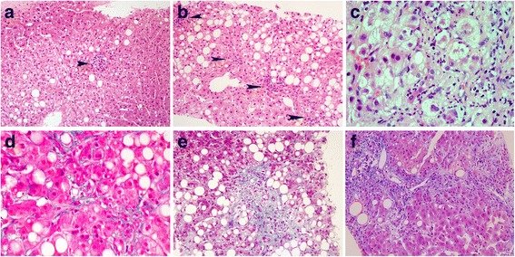
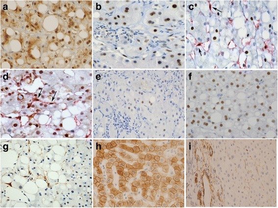
그림 출처: PubMed Central 공개 논문 (PMC5883355)
Conflict of interest statement
ETHICS APPROVAL AND CONSENT TO PARTICIPATE: All human liver tissue specimens were obtained during diagnostic and therapeutic procedures. Consent to participate was not required because this was a retrospective study of formalin-fixed, paraffin-embedded tissues obtained after pathological diagnosis. All samples were anonymized before initiating the study. The retrospective study protocol was reviewed and approved by the Institutional Review Board of the Seoul National University Boramae Hospital with the condition of anonymization (IRB No. 26–2016-12/022). The study was conducted in accordance with the principles of the Declaration of Helsinki. CONSENT FOR PUBLICATION: Not applicable COMPETING INTERESTS: The authors declare no conflicts of interest. PUBLISHER’S NOTE: Springer Nature remains neutral with regard to jurisdictional claims in published maps and institutional affiliations.
그 밖의 정보
- 출판 형태
- Journal Article
- 저널
- Diagnostic pathology · eISSN 1746-1596
- 전자 출판
- 2018-04-03
- MeSH
- Adult · Child · Disease Progression · Estrogen Receptor alpha / biosynthesis · Extracellular Signal-Regulated MAP Kinases · Fatty Liver / pathology · Female · Humans · Male · Middle Aged · Retrospective Studies · STAT3 Transcription Factor / biosynthesis
- Substances
- ESR1 protein, human · Estrogen Receptor alpha · STAT3 Transcription Factor · STAT3 protein, human · Extracellular Signal-Regulated MAP Kinases
- 연구비
- the Basic Science Research Program of the National Research Foundation of Korea, the Ministry of Education 2016R1D1A1B01010316
- 참고문헌
- 55편