SCI 발표논문

SCI 발표논문

← SCI 발표논문 목록

PLoS One. 2018;13(8):e0201692.

HL156A, a novel pharmacological agent with potent adenosine-monophosphate-activated protein kinase (AMPK) activator activity ameliorates renal fibrosis in a rat unilateral ureteral obstruction model.

, , , , , , , , , , ,

제1저자 Bodokhsuren · 교신저자 오국환

소속 (7)
  1. Biomedical Research Institute, Seoul National University Hospital, Seoul, Korea.
  2. Department of Internal Medicine, Seoul National University College of Medicine, Seoul, Korea.
  3. Renal Division, Department of Internal Medicine, Seoul National University Hospital, Seoul, Korea.
  4. Department of Clinical Pharmacology and Therapeutics, Seoul National University College of Medicine and Hospital, Seoul, Korea.
  5. Department of Internal Medicine, Seoul National University Boramae Medical Center, Seoul, Korea.
  6. Department of Internal Medicine, Dongkuk University, Kyungju, Korea.
  7. Transplantation Research Institute, Seoul National University Hospital, Seoul, Korea.

Abstract

BACKGROUND Renal fibrosis is characterized by excessive production and deposition of extracellular matrix (ECM), which leads to progressive renal failure. Adenosine-monophosphate-activated protein kinase (AMPK) is a highly conserved kinase that plays a key role in Smad-3 signaling. Here, we examined the effect of a novel AMPK activator, HL156A, on the inhibition of renal fibrosis in in vivo and in vitro models.

METHODS Unilateral ureteral obstruction (UUO) was induced in male Wistar rats. Rats with UUO were administered HL156A (20mg/kg/day), and then the kidneys were harvested 10 days after ligation for further analysis.

RESULTS In the rat UUO model, HL156A attenuated ECM protein deposition. After HL156A treatment, expressions of TGF-β1, p-Smad3, α-SMA, fibronectin, and type IV collagen were suppressed, and E-cadherin expression was up-regulated. In the in vitro experiment, NRK52E cells were treated with HL156A before TGF-β1 stimulation. The inhibitory effects of HL156A upon the signaling pathways and markers of the epithelial-to-mesenchymal transition (EMT) were analyzed. In TGF-β1-treated NRK-52E cells, HL156A co-treatment inhibited the TGF-β1-induced Smad3 signaling pathway and EMT markers.

CONCLUSION Taken together, the above findings suggest that HL156A, a novel AMPK activator, ameliorates renal fibrosis in vivo and in vitro.

Figures

Fig 1
Fig 1 Comparison of AMPK activator activities. AMPK activities induced by various AMPK activator molecules were determined on rat renal proximal tubular (NRK-52E) cells. The AMPK activator activities of 5-aminoimidazole-4-carboxamide-1-β-D-ribofuranoside (AICAR), metformin, and HL156A were compared at various concentrations (0, 10, 30, and 50 μM, respectively). AMPK activator activity was determined by AMPKαThr172 phosphorylation and was measured using an ELISA kit (Invitrogen Corporation). Experiments were repeated at three times. +: P <0.05 vs. AICAR, ‡: P <0.05 vs. metformin at the same concentration.
Fig 2
Fig 2 The effect of HL156A treatment on UUO. In a preliminary experiment, a unilateral ureteral obstruction (UUO) model was induced in adult male Wistar rats. Obstructed kidneys were harvested 10 days after surgery. (A) Each ligated kidney was compared with its contralateral (non-ligated) kidney after Masson’s trichrome staining. (B) To find the optimal dose of HL156A, the rats were subjected to unilateral ureter ligation and then treated with various doses of HL156A (0, 10, and 20 mg/kg/day for 10 days) before comparative analyses were performed. Experiments were repeated at two times with N = 4 each time. Treatment with 20mg/kg of HL156A markedly ameliorated the renal injury from UUO.
Fig 3
Fig 3 HL156A ameliorated histopathological changes associated with UUO. A unilateral ureteral obstruction (UUO) model was induced in adult male Wistar rats. Rats with UUO were administered HL156A (20 mg/kg/day) or saline one day before the ligation surgery and daily thereafter. Obstructed kidneys were harvested 10 days after surgery. (A) Masson’s trichrome shows that UUO induced severe renal fibrosis, which was significantly ameliorated by HL156A. (B) Quantification of renal injury score based on the Masson’s trichrome staining of the kidneys. (C) Immunohistochemical staining shows UUO down-regulated p-AMPK, thereby inducing an increase in α-SMA and a down-regulation of E-cadherin, both of which are hallmarks of EMT. UUO increased expression of extracellular matrix (ECM) molecules, such as fibronectin and type IV collagen. HL156A treatment, which recovered down-regulated p-AMPK, significantly diminished the above changes induced by UUO. Experiments were repeated at two times with N = 7 each time. * P <0.05 vs. Sham, #P <0.05 between UUO and UUO + HL156A.
Fig 4
Fig 4 Effects of HL156A treatment on mRNA and protein expression. mRNA and protein expressions of various markers were analyzed from the animal experiment. (A) Reverse transcription PCR was used to analyze mRNA expression levels of AMPKα1, AMPKα2, TGF- β1, Smad3, α-SMA, fibronectin, E-cadherin, and type IV collagen. HL156A treatment recovered the down-regulations of AMPKα1 and AMPKα2 induced by UUO. (B) Western blot analysis was used to measure phosphorylated AMPKα, total AMPK, TGF- β1, Smad3, α-SMA, fibronectin, E-cadherin, and type IV collagen expression levels from kidney tissue. (C) Western blot analysis of protein bands quantified using image J and normalized to β-actin. Experiments were repeated two times with N = 7 at each time. *P <0.05 vs. sham, #P <0.05 between UUO and UUO + HL156A.
Fig 5
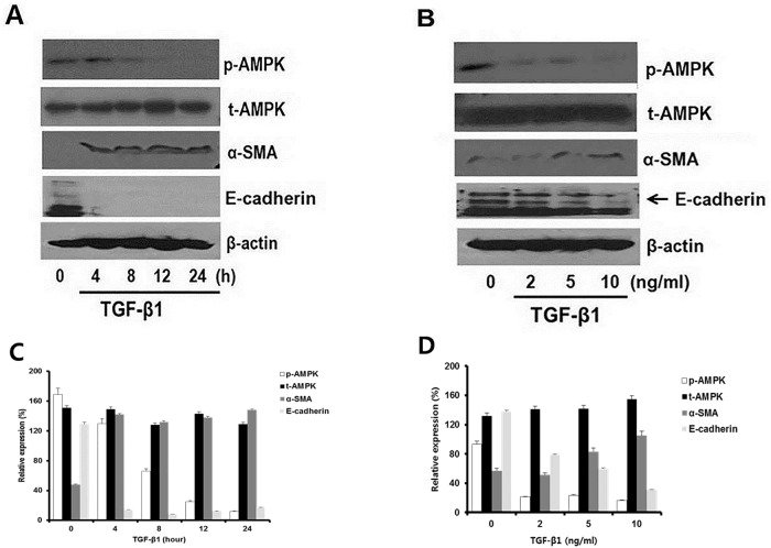
Fig 5 Pilot study to estimate the optimal dose and duration of TGF-β1 treatment. Cultured rat proximal tubular NRK-52E cells were incubated in the different concentrations TGF-β1 and various exposure time. (A) NRK-52E cells were treated with TGF-β1 at different concentrations (0, 2, 5, and 10 ng/ml) for 24 hours and were subjected to western blot analysis to evaluate phosphorylation of AMPK, total AMPK, α-SMA, E-cadherin, and β-actin. (B) NRK-52E cells were exposed to TGF-β1 at various exposure times (0, 4, 8, 12, and 24 hours), and AMPK, α-SMA, and E-cadherin expressions were evaluated. The results showed that 10 ng/ml of TGF-β1 for 24 hours were the optimal dose and time for our further experiments. (C, D) Western blot analysis quantified protein bands by using image J and normalized to β-actin. Experiments were repeated three times.
Fig 6
Fig 6 The effect of HL156A on TGF-β1-treated NRK52E cells. NRK-52E cells, cultured rat proximal cells, were incubated in the presence or absence of HL156A 30 μM for 24 hours. Then, these cells were stimulated with TGF-β1 (10 ng/mL) for 24 hours before harvesting. Compared with TGF-β1 treatment alone, (A) HL156A co-treatment up-regulated AMPKα1, AMPKα2, and E-cadherin expressions. HL156A down-regulated Smad3, α-SMA, fibronectin, and type IV collagen. This was confirmed by mRNA expression levels of AMPKα1, AMPKα2, TGF-β1, Smad3, α-SMA, fibronectin, E-cadherin, and type IV collagen. (B) Representative Western blots of phosphorylated AMPKα, total AMPK TGF-β1, Smad3, α-SMA, fibronectin, E-cadherin, and type IV collagen expression levels from the NRK-52E cells. (C) Western blot analysis of protein bands quantified using image J and normalized to β-actin. Experiments were repeated three times. *P <0.05 vs. vehicle, #P < 0.05 between TGF-β1 and TGF-β1 + HL156A.
Fig 7
Fig 7 The expression of inflammatory cytokine. (A) Immunohistochemistry (IHC) staining showed expression of MCP-1 (brown). This revealed that HL156A inhibited MCP-1, which was induced by UUO group. (B) qRT-PCR and western blot data showed that HL156A treatment improved the MCP-1 under the UUO group. (C) HL156A treatment induced of MCP-1 from NRK52E cells. Experiments were repeated two times. *P <0.05 vs. Sham or vehicle, #P < 0.05 between TGF-β1 and TGF-β1 + HL156A.
Fig 8
Fig 8 Immunofluorescence analysis of AMPK and Smad3 pathways. NRK52E cells were treated with TGF-β1 (10 ng/ml) with or without HL156A (30 μM) for 24 hours. (A) Immunofluorescence staining showed expression of p-AMPK (red), p-Smad3 (red), E-cadherin (red), α-SMA (green), fibronectin (green), and type IV collagen (green). This revealed that HL156A, by stimulating p-AMPK, inhibited p-Smad3, α-SMA, fibronectin, and type IV collagen, which were all induced by TGF-β1 treatment. HL156A co-treatment also restored E-cadherin expression that was suppressed under TGF-β1 treatment. (B) Immunofluorescence analysis quantified using image J. Experiments were repeated three times. *P <0.05 vs. Vehicle, #P < 0.05 between TGF-β1 and TGF-β1 + HL156A.

그림 출처: PubMed Central 공개 논문 (PMC6116936)

Conflict of interest statement

This research was funded by Hanall Biophama Co., Korea. No participating authors are employees, or consultants of the company. Besides, this does not alter our adherence to PLOS ONE policies on sharing data and materials.

그 밖의 정보
출판 형태
Journal Article, Research Support, Non-U.S. Gov't
저널
PloS one · eISSN 1932-6203
전자 출판
2018-08-30
MeSH
AMP-Activated Protein Kinases / metabolism · Animals · Apoptosis / drug effects · Cell Line · Disease Models, Animal · Dose-Response Relationship, Drug · Enzyme Activation / drug effects · Fibrosis · Guanidines / pharmacology / therapeutic use · Kidney / drug effects / metabolism / pathology · Pyrrolidines / pharmacology / therapeutic use · Rats · Rats, Wistar · Signal Transduction / drug effects · Smad3 Protein / metabolism · Time Factors · Transforming Growth Factor beta1 / metabolism · Ureteral Obstruction / pathology
Substances
Guanidines · HL156A · Pyrrolidines · Smad3 Protein · Transforming Growth Factor beta1 · AMP-Activated Protein Kinases
참고문헌
35편