Biomedical Research Institute, Seoul National University Hospital, Seoul, Korea.
Molecular Medicine & Biopharmaceutical Sciences, Seoul National University, Seoul 03080, Republic of Korea.
Division of Cardiology, Cardiovascular Center, Seoul National University Bundang Hospital, Seongnam, Korea.
Korean Medical Science Research Center for Healthy-Aging, Graduate Training Program of Korean Medicine for Healthy-Aging, Pusan National University, Yangsan, Republic of Korea.
UMR-1132 INSERM and University Paris Diderot, Sorbonne Paris Cité, Hôpital Lariboisiére, Paris, France.
Department of Internal Medicine, Seoul National University College of Medicine, Seoul, Republic of Korea.
Abstract
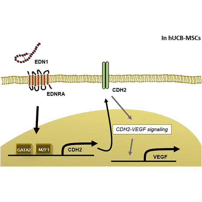
In our previous study, we identified differences in the levels of CDH2 and vascular endothelial growth factor (VEGF) between effective and ineffective clones of human umbilical cord blood (hUCB) mesenchymal stem cells (MSCs), with regard to the infarcted rat myocardium. In this study, we compared gene expression profiles between the effective and ineffective clones and identified that endothelin-1 (EDN1) is enriched in the effective clone. In the mechanistic analyses, EDN1 significantly increased expression of CDH2 and VEGF through endothelin receptor A (EDNRA), which was prevented by EDNRA blocker, BQ123. To decipher how EDN1 induced gene expression of CDH2, we performed a promoter activity assay and identified GATA2 and MZF1 as inducers of CDH2. EDN1 significantly enhanced the promoter activity of the CDH2 gene, which was obliterated by the deletion or point mutation at GATA2 or MZF1 binding sequence. Next, therapeutic efficacy of EDN1-priming of hUCB-MSCs was tested in a rat myocardial infarction (MI) model. EDN1-primed MSCs were superior to naive MSCs at 8 weeks after MI in improving myocardial contractility (p < 0.05), reducing fibrosis area (p < 0.05), increasing engraftment efficiency (p < 0.05), and improving capillary density (p < 0.05). In conclusion, EDN1 induces CDH2 and VEGF expression in hUCB-MSCs, leading to the improved therapeutic efficacy in rat MI, suggesting that EDN1 is a potential priming agent for MSCs in regenerative medicine.
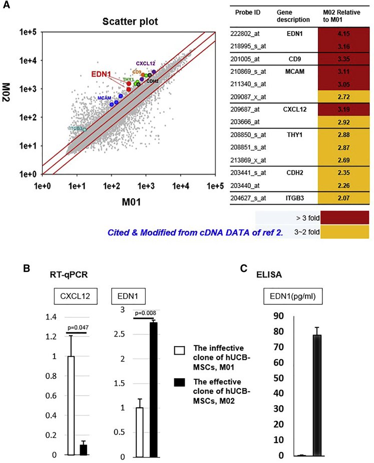
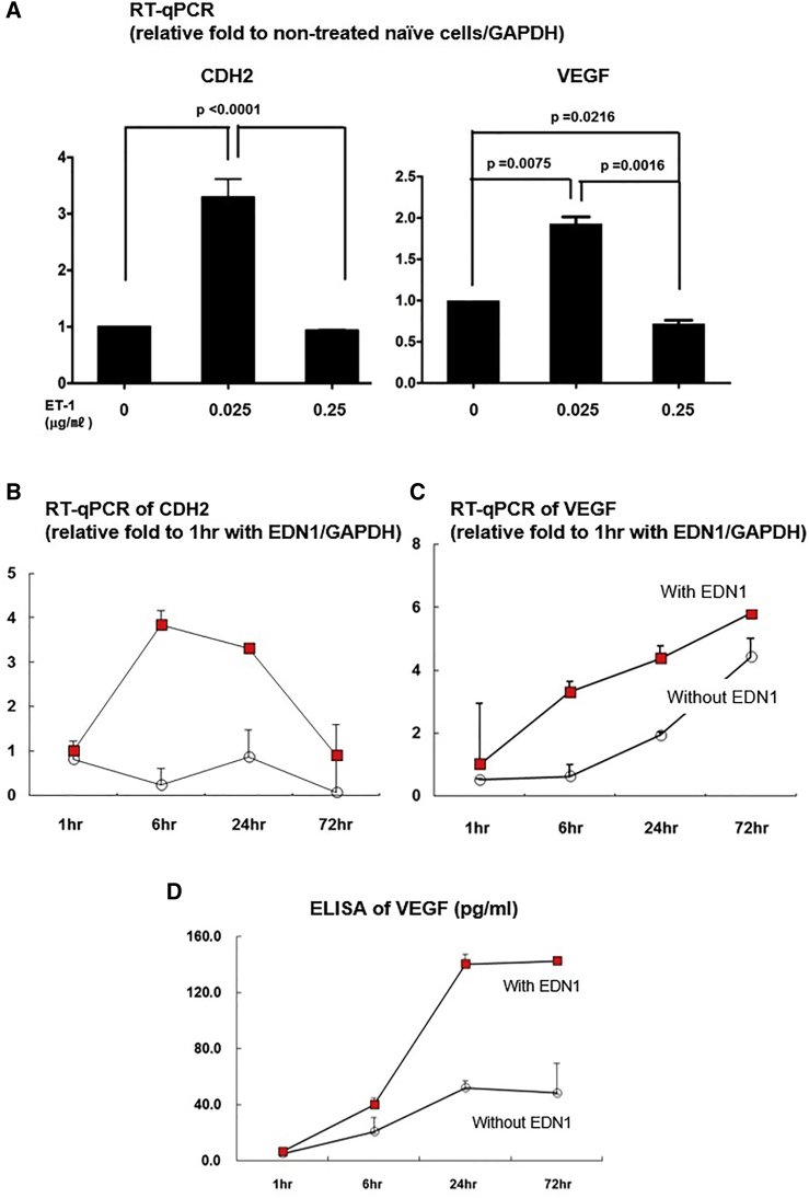
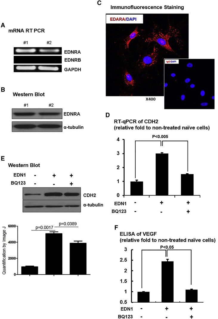
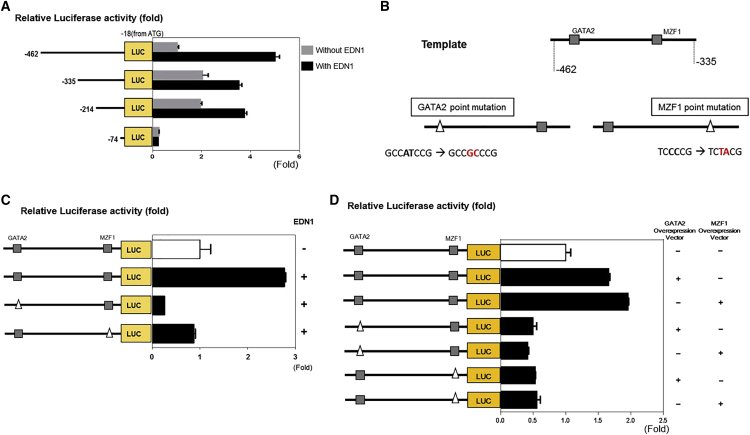
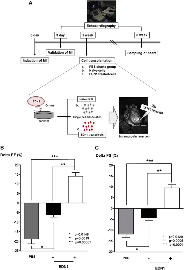
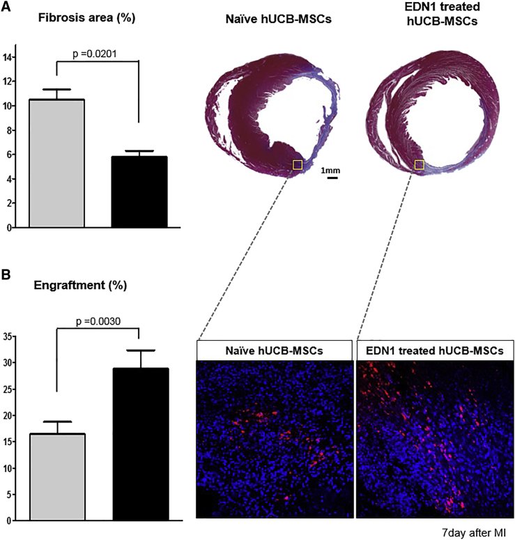
Figure 1Screening for a Factor to Enhance the Regenerative Capability of hUCB-MSCs (A) Scheme of comparison of the different gene expression profiles of the effective hUCB-MSCs, M02, and the ineffective ones, M01. Scatterplot of whole gene expression profiles of M02 and M01 from our previous report.2 Screened seven genes represented according to expression level of M02 relative to M01 are shown. First, surface and secretory molecules were selected and their statistically significant differences in expression were identified (p ≤ 0.05). Among them, we selected secretory factors, EDN1and CXCL12, for application as priming factors. (B) Expression of EDN1 and CXCL12 mRNA were confirmed using qRT-PCR. EDN1 mRNA showed significantly higher expression in the effective clone of hUCB-MSCs than in the ineffective one. The levels of CXCL12 mRNA did not change significantly. (C) ELISA showed higher EDN1 secretion from the effective clone.Figure 2EDN1 Upregulates Expression of CDH2 and VEGF (A) qRT-PCR to determine the levels of CDH2 and VEGF mRNA. Transcription of CDH2 and VEGF increased with 0.025 μg/mL EDN1 compared with 0 and 0.25 μg/mL EDN1. (B–D) Optimization of EDN1 treatment time. (B) qRT-PCR to determine the levels of CDH2 is shown. Expression of CDH2 mRNA peaked at 6 h. (C) qRT-PCR to determine the levels of VEGF is shown. VEGF expression continues to increase from 1 to 72 h following EDN1 treatment. (D) ELISA to determine the levels of VEGF protein is shown. Secreted VEGF increased from 6 h and peaked at 24∼72 h. Taken together, the optimal condition of EDN1 treatment of cells is 0.025 μg/mL for 24 h.Figure 3EDN1 Induced CDH2 and VEGF Levels through EDNRA Expression of EDN1 receptors was assessed at the mRNA and protein levels in ineffective hUCB-MSCs. Only EDNRA was expressed in ineffective hUCB-MSCs. (A) RT-PCR to determine EDNRA and EDNRB mRNA is shown. (B) Western blot of EDNRA is shown. (C) Immunofluorescence staining of EDNRA is shown. (D) qRT-PCR to determine the levels of CDH2 mRNA is shown. (E) Western blot to determine the levels of CDH2 protein is shown. An EDNRA blocker, BQ123, suppressed upregulation of CDH2 mRNA and protein under EDN1 treatment. (F) ELISA to determine the levels of VEGF protein is shown. BQ123 reduced levels of secreted VEGF following treatment with EDN1. Overall, EDNRA mediates induction of the CDH2 and VEGF by EDN1.Figure 4GATA2 and MZF1 Are Transcription Factors of CDH2 Induction by EDN1 (A) Luciferase reporter assay to determine activity of the CDH2 promoter region (the −462- to −18-bp fragment from the ATG) and fragments of the promoter sequence (bases −335 to −18, −214 to −18, and −74 to −18). EDN1 significantly enhanced the promoter activity (−462- to −18-bp fragment; p < 0.05). Deletion of the sequence between −462 and −335 bp retarded this effect (−462 to −18 bp versus −335 to −18; p < 0.05). (B) The sequence from −462 to −335 bp contained one GATA2 and one MZF1 binding sequence. Putative GATA2 and MZF1 binding regions were identified using the TFSEARCH database v1.3, and point mutagenesis was carried out. (C) Luciferase reporter assay to determine the promoter activity of the −462- to −335-bp fragment with and without point mutations. The promoter activity of the −462- to −335-bp region was increased by EDN1 treatment (p < 0.05) but was obliterated by point mutations at either GATA2 or MZF1 site (p < 0.05). (D) Luciferase reporter assay to determine the promoter activity of the −462- to −335-bp fragment with and without point mutations in cells overexpressing GATA2 or MZF1 is shown. The promoter activity of the −462- to −335-bp region was increased by co-expression of GATA2 or MZF1 (p < 0.05). Reduced activity was observed in each point mutant (ΔGATA2-MZF1 or ΔMZF1-GATA2; no significant difference).Figure 5Transplantation of EDN1-Primed hUCB-MSCs Improved Myocardial Regeneration in a Rat Myocardial Infarction (MI) Model (A) Scheme for cell transplantation in MI. EDN1-primed and naive ineffective hUCB-MSCs were transplanted in infarcted heart at 1 week after induction of MI. (B) 8 weeks after induction of MI, delta EF in the EDN1-treated hUCB-MSC group was improved significantly compared to that in the naive hUCB-MSCs or PBS-injected groups (p < 0.05). (C) 8 weeks after induction of MI, delta FS was significantly improved in the EDN1 group compared to the control MSCs or PBS-injected groups (p < 0.05). *p = 0.01, PBS versus naive MSCs; **p = 0.001, naive MSCs versus EDN1-primed MSCs; and ***p = 0.00007, PBS versus EDN1-primed MSCs in delta EF. *p = 0.01, PBS versus naive MSCs; **p = 0.0005, naive MSCs versus EDN1-primed MSCs; and ***p = 0.0001, PBS versus EDN1 primed MSCs in delta FS (n = 7 in each group).Figure 6EDN1-Primed hUCB-MSCs Survive Longer after Implantation and Reduce Infarct Scar in the Rat MI Model (A) Masson’s trichrome staining of infarcted rat hearts and quantitative analysis of the fibrosis area (blue). The fibrosis area was significantly reduced in the EDN1-treated hUCB-MSC group compared with the naive hUCB-MSCs group (p = 0.02). (B) Analysis of the engraftment using DiI labeling of transplanted hUCB-MSCs is shown. The intensity of DiI-labeled cells was higher after transplantation of EDN1-treated hUCB-MSCs than after transplantation of naive hUCB-MSCs (p = 0.003).
그림 출처: PubMed Central 공개 논문 (PMC6545354)
그 밖의 정보
출판 형태
Journal Article
저널
Molecular therapy. Methods & clinical development · ISSN 2329-0501