
PLoS One. 2017;12(9):e0185501.
Risk factors for early metachronous tumor development after endoscopic resection for early gastric cancer.
제1저자 박재용 · 교신저자 김상균
소속 (3)
- Department of Internal Medicine, Chung-Ang University Hospital, Seoul, Korea.
- Department of Internal Medicine and Liver Research Institute, Seoul National University College of Medicine, Seoul, Korea.
- Department of Internal Medicine, Healthcare Research Institute, Seoul National University Hospital Healthcare System Gangnam Center, Seoul, Korea.
Abstract
BACKGROUND Metachronous gastric tumor (MGT) is one of major concerns after endoscopic submucosal dissection (ESD) for early gastric cancer (EGC). Optimal follow-up strategy has not been yet well-established. The aim of this study was to identify the different clinical features of the patients according to the time interval to development of MGT.
METHODS Among 1,780 consecutive patients with EGC who underwent ESD between 2005 and 2014, 115 patients with MGT were retrospectively reviewed. MGT was defined as secondary gastric cancer or dysplasia detected > 1 year after initial ESD. Clinicopathological factors associated with early development of MGT were evaluated.
RESULTS The median interval to development of MGT was 37 months. In univariate analysis, the median interval to MGT was shorter if EGC lesion was non-elevated type (39.4 vs 57.0 months, p = 0.011), or synchronous primary lesion was absent (39.8 vs 51.4 months, p = 0.050). In multivariate Cox's proportional hazards analysis, the hazard ratios for early occurrence of MGT were 1.966 (95% CI: 1.141-3.386, p = 0.015) and 1.911 (95% CI: 1.163-3.141, p = 0.011), respectively. There was no significant difference in overall survival after diagnosis of MGT between the early occurrence group and the late occurrence group.
CONCLUSIONS Non-elevated gross type and absence of synchronous gastric tumor were independent risk factors for early development of MGT. Meticulous endoscopic inspection is especially important for the detection of MGT during the early follow-up period in patients with these initial tumor characteristics.
Figures

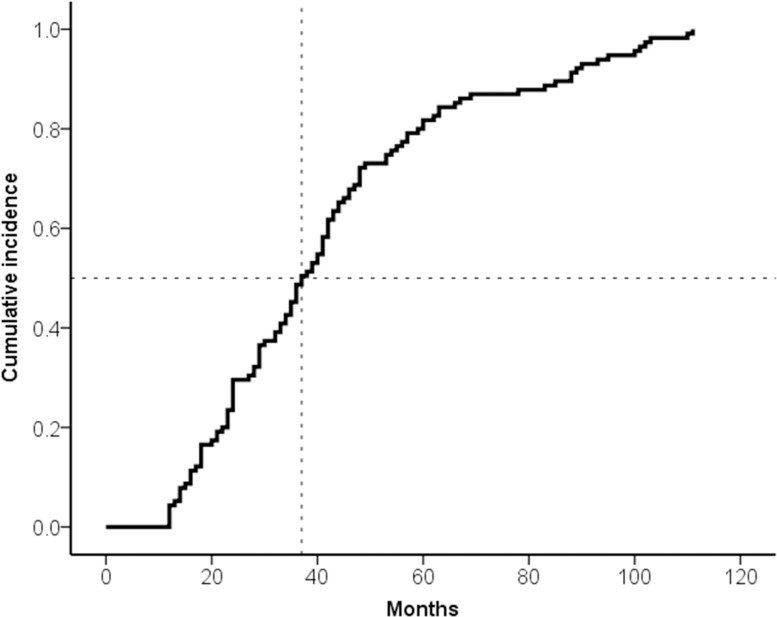
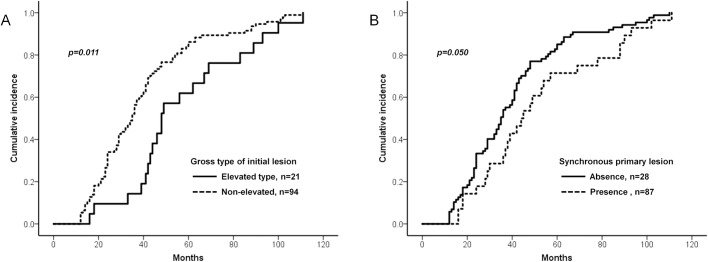
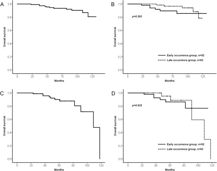
그림 출처: PubMed Central 공개 논문 (PMC5614623)
Conflict of interest statement
Competing Interests:The authors have declared that no competing interests exist.
그 밖의 정보
- 출판 형태
- Journal Article
- 저널
- PloS one · eISSN 1932-6203
- 전자 출판
- 2017-09-26
- MeSH
- Aged · Endoscopy / methods · Female · Gastroscopy / methods · Helicobacter Infections / complications / drug therapy / microbiology · Helicobacter pylori / isolation & purification · Humans · Male · Middle Aged · Retrospective Studies · Risk Factors · Stomach Neoplasms / complications / pathology / surgery · Survival Analysis
- 참고문헌
- 33편