Strategic Center of Cell and Bio Therapy for Heart, Diabetes & Cancer, Biomedical Research Institute, Seoul National University Hospital, Seoul, 03080, Republic of Korea.
Clinical Research Team, SK Chemical, Life Science Biz., Seongnam-si, Gyeonggi-do, 13494, Republic of Korea.
Skin Research Division, AMOREPACIFIC Corp. R&D Unit, Yongin, -si, Gyeonggi-do, Republic of Korea.
Departments of Dermatology, Chung-Ang University College of Medicine, Seoul, Republic of Korea.
Department of Convergence Medicine, Pusan National University School of Medicine, Yangsan, 50612, Korea.
Strategic Center of Cell and Bio Therapy for Heart, Diabetes & Cancer, Biomedical Research Institute, Seoul National University Hospital, Seoul, 03080, Republic of Korea. usahyosoo@gmail.com.
Molecular Medicine and Biopharmaceutical Sciences, Graduate School of Convergence Science and Technology, Seoul National University, Seoul, 03080, Republic of Korea. usahyosoo@gmail.com.
Cardiovascular Center & Department of Internal Medicine, Seoul National University Hospital, Seoul, Republic of Korea. usahyosoo@gmail.com.
Abstract
BACKGROUND The human skin-derived precursors (SKPs) are a good cell source for regeneration. However, the isolation of SKP from human skin is limited. To overcome this drawback, we hypothesized that the component of plant stem cells could convert human fibroblasts to SKPs.
METHODS Human dermal fibroblasts were treated with shikimic acid, a major component of Sequoiadendron giganteum callus extract. The characteristics of these reprogrammed cells were analyzed by qPCR, western blot, colony-forming assay, and immunofluorescence staining. Artificial human skin was used for CO2 laser-induced wound experiments. Human tissues were analyzed by immunohistochemistry.
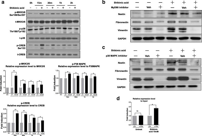
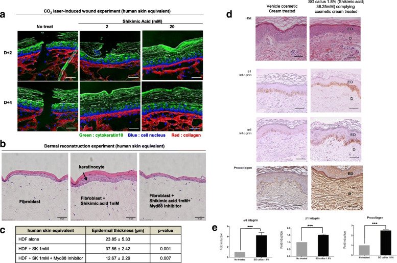
RESULTS The reprogrammed cells expressed nestin (a neural precursor-specific protein), fibronectin, and vimentin and could differentiate into the ectodermal and mesodermal lineage. Nestin expression was induced by shikimic acid through the mannose receptor and subsequent MYD88 activation, leading to P38 phosphorylation and then CREB binding to the nestin gene promoter. Finally, we confirmed that shikimic acid facilitated the healing of cut injury and enhanced dermal reconstruction in a human artificial skin model. Moreover, in a clinical study with healthy volunteers, plant callus extracts increased the expression of stem cell markers in the basal layer of the epidermis and collagen deposit in the dermis.
CONCLUSIONS These results indicate that shikimic acid is an effective agent for tissue regeneration.
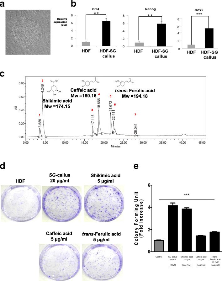
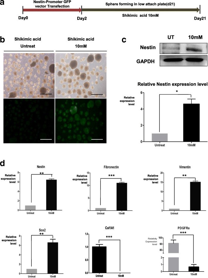
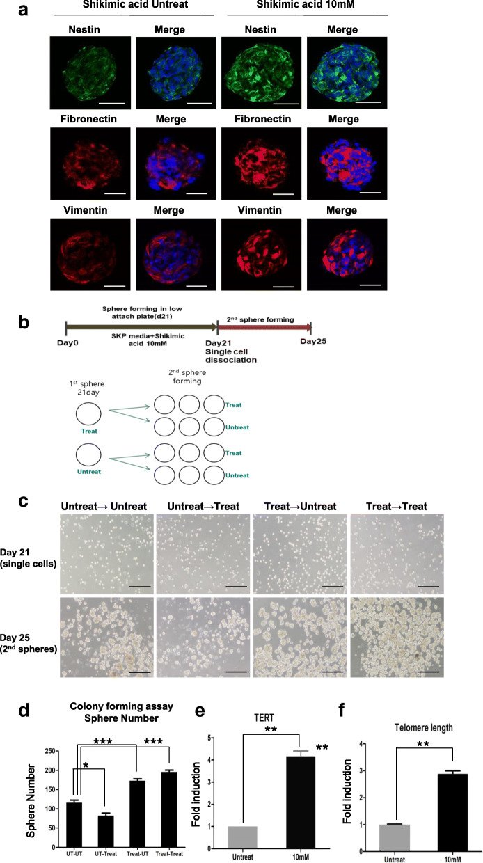
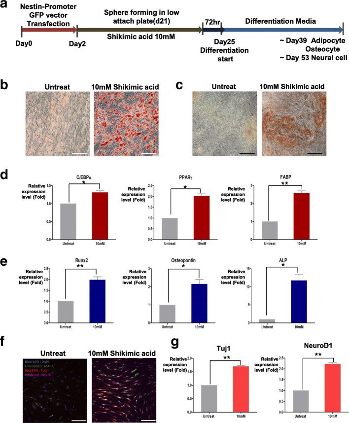
Fig. 1SG-callus extract and its major component, shikimic acid, induce stemness in human fibroblast and expression of pluripotent stem cell markers. a Bright-field images of HDF after treatment with SG callus extract (500 μg/ml) for 7 days. Scale bar = 500 μm. b RNA expression of OCT4, NANOG, and Sox2 were analyzed by qRT-PCR. It was normalized to 18s RNA (N = 3 in each group, **p < 0.01). c HPLC chromatogram of the SG-callus extract detected at the UV wavelength of 230 nm. The peak (RT = 4.248 min) is assigned to the shikimic acid. The peak (RT = 18.666 min) is assigned to the caffeic acid. The peak (RT = 21.672 min) is assigned to the trans-ferulic acid. d A colony formation assay was performed on naive HDF and HDF treated with SG-callus extract or shikimic acid, caffeic acid, and trans-ferulic acid at 10 days after treatment. Scale bar = 500 μm. Representative pictures are shown. e Relative stained colonies are quantified (N = 3 in each group, *p < 0.05, ***p < 0.001 compared with the control)Fig. 2NeuSKPCs are induced by treatment of shikimic acid. a Scheme of generation of NeuSKPCs. b Morphology of NeuSKPCs. Nestin promoter GFP was activated in the shikimic acid-treated groups (scale bar = 500 μm). c Western blot analysis was conducted using an anti-nestin antibody (upper panel) in shikimic acid untreated or treated spheroids, and the quantified result is shown in the lower panel (*p < 0.05). d qRT-PCR analysis of SKP-associated genes, nestin, fibronectin, vimentin, pluripotency-associated gene, Sox2, fibroblast-specific genes, Col1A1, and PDGFRa. It was normalized to 18s RNA (N = 3 in each group, **p < 0.01, ***p < 0.001 compared with the control)Fig. 3Characterization of shikimic acid-induced NeuSKPCs. a Immunofluorescence of SKP markers including anti-nestin (green), anti-fibronectin, and anti-vimentin (red) in shikimic acid-induced NeuSKP on day 21. DAPI was used to identify the nucleus of the cells (scale bar = 50 μm). b Scheme of colony-forming assay (secondary sphere formation). c Bright-field images of secondary NeuSKPCs with or without shikimic acid for 4 days (scale bar = 500 μm). d The number of secondary spheres from matured NeuSKPCs (N = 5 in each group, *p < 0.05, ***p < 0.001). e qRT-PCR analysis of TERT in matured NeuSKPCs (10 mM shikimic acid was treated for 21 days, **p < 0.01 compared with the untreated control). f Comparison of the average telomere length ration between shikimic acid untreated or treated (**p < 0.01 compared with the untreated control)Fig. 4Shikimic acid-induced NeuSKPCs can differentiate into ectodermal and mesodermal lineage. a Scheme of differentiation for NeuSKPCs. b Lipid drops of adipogenic differentiated cells were stained by Oil red O (upper panel, scale bar = 100 μm). c Accumulated calcium of osteogenically differentiated cells were stained by von Kossa staining (scale bar = 200 μm). d qRT-PCR analysis of adipocyte-specific genes, CEBP, PPAR, and FABP (N = 3 in each group; ns, not significant, *p < 0.05, **p < 0.01). e qRT-PCR analysis of osteocyte-specific genes, Runx2, osteopontin, and ALP (N = 3 in each group, *p < 0.05, **p < 0.01). f Differentiated neural cells were stained with MAP2 (green), Tuj1(red), and NeuN (magenta) (scale bar = 100 μm). g qRT-PCR analysis of neuron-specific genes, Tuj1, and NeuroD1 (N = 3 in each group, **p < 0.01)Fig. 5Shikimic activates nestin expression via the MyD88/p38/CREB signaling pathway. a After the designated incubation time with shikimic acid, the phosphorylation level of MAPK-kinase (MKK)3/6, p38 MAPK, and CREB was determined (upper). Quantification of the obtained results (N = 3 in each group, *p < 0.05, **p < 0.01, ***p < 0.001 compared with the control) (lower). b Western blot analysis of nestin, fibronectin, and vimentin after treatment of MyD88 inhibitor and shikimic acid. c Western blot analysis of nestin, fibronectin, and vimentin after treatment of p38 inhibitor and shikimic acid. d Chromatin immunoprecipitation (ChIP) assay on the promoter of nestin. Phosphor-CREB antibody was used for the ChIP assay (N = 3 in each group, *p < 0.05)Fig. 6Shikimic acid facilitates wound healing and enhances dermal reconstruction in the artificial human skin ex-vivo model. a Representative fluorescent immuno-histologic staining of cytokeratin (green) in the epidermis and cell nucleus (blue) in the basement membrane and collagen (red) in the dermis from human skin equivalent tissue. Tissue was frozen in OCT and stained by the immunofluorescence method. Data are representative of N = 3. Scale bar = 10 μm. b The dermal layers were seeded with 3 different fibroblasts: Non-treated fibroblast (control), fibroblast treated with shikimic acid, or fibroblast treated with shikimic acid plus MyD88 inhibitor. Keratinocytes were seeded over the dermal layer. This skin equivalent was cultured in the air-liquid interface for 8 days. On 8 days, the paraffin sections were stained for hematoxylin and eosin (scale bar = 50 μm). c The quantification of epidermal thickness (N = 3 in each group compared with control HDF, p < 0.001 or p < 0.007). dSG-callus extract (36.25 mM shikimic acid) increased the expression level of β1 integrin and α6 integrin that are stem cell markers at the basal layer of the epidermis. It also increased collagen amounts in the dermis (brown color in immunostaining) (scale bar = 100 μm). eN = 8, treated group compared with vehicle cream treated group, ***p < 0.001. ED, epidermis; D, dermis
그림 출처: PubMed Central 공개 논문 (PMC8196440)
Conflict of interest statement
The authors declare that they have no competing interests.