Institute of Allergy and Clinical Immunology, Seoul National University Medical Research Center, 103 Daehak-ro, Jongno-gu, Seoul, 03080, Korea.
Department of Translational Medicine, Seoul National University College of Medicine, 103 Daehak-ro, Jongno-gu, Seoul, 03080, Korea.
Department of Medical Science, Seoul National University College of Medicine, 103 Daehak-ro, Jongno-gu, Seoul, 03080, Korea.
Department of Internal Medicine, Seoul National University College of Medicine, 103 Daehak-ro, Jongno-gu, Seoul, 03080, Korea.
Institute of Allergy and Clinical Immunology, Seoul National University Medical Research Center, 103 Daehak-ro, Jongno-gu, Seoul, 03080, Korea. helenmed@snu.ac.kr.
Department of Translational Medicine, Seoul National University College of Medicine, 103 Daehak-ro, Jongno-gu, Seoul, 03080, Korea. helenmed@snu.ac.kr.
Department of Internal Medicine, Seoul National University College of Medicine, 103 Daehak-ro, Jongno-gu, Seoul, 03080, Korea. helenmed@snu.ac.kr.
Abstract
Mesenchymal stem cells (MSCs) possess immunomodulatory properties that have therapeutic potential for the treatment of inflammatory diseases. This study investigates the effects of direct MSC administration on asthmatic airways. Umbilical cord MSCs (ucMSCs) were intratracheally administered to six-week-old female BALB/c mice sensitized and challenged with ovalbumin; airway hyperresponsiveness (AHR), analyses of airway inflammatory cells, lung histology, flow cytometry, and quantitative real-time PCR were performed. Furthermore, ex vivo and in vitro experiments were performed to assess the effects of ucMSC on M2 activation. Intratracheally administered ucMSCs decreased degree of airway resistance and the number of inflammatory cells such as T helper 2 (Th2) cells, type 2 innate lymphoid cells (ILC2), and macrophages in the murine asthma model. Particularly, MHCII and CD86 expression diminished in dendritic cells and alveolar macrophages (AMs) following ucMSC treatment. SiglecF+CD11c+CD11b- AMs show a negative correlation with type II inflammatory cells including Th2 cells, ILC2, and eosinophils in asthmatic mice and were restored following intratracheal ucMSCs treatment. In addition, ucMSCs decreased the macrophage polarization to M2, particularly M2a. The expression levels of markers associated with M2 polarization and Th2 inflammation were also decreased. ucMSC reduced Il-12 and Tnfa expression as well as that of M2 markers such as Cd206 and Retnla ex vivo. Furthermore, the in vitro study using IL-4 treated macrophages confirmed that both direct and indirect MSC treatment significantly reduced the expression of Il-5 and Il-13. In conclusion, ucMSCs appear to suppress type II inflammation by regulating lung macrophages via soluble mediators.
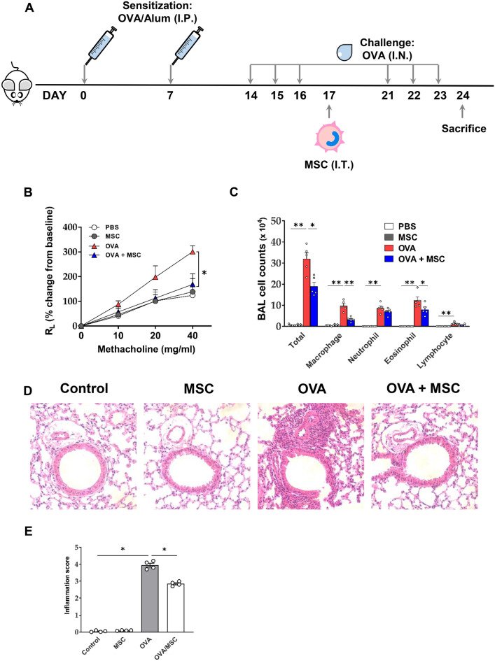
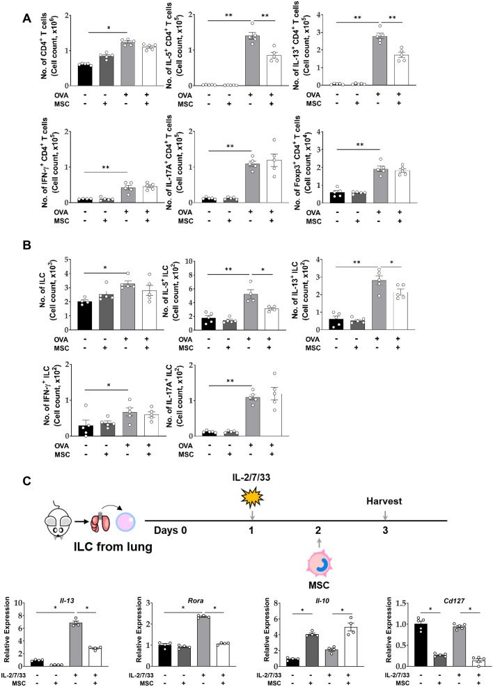
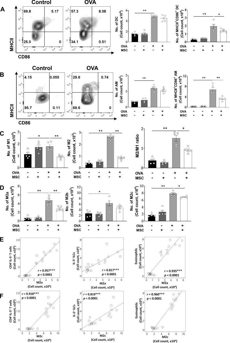
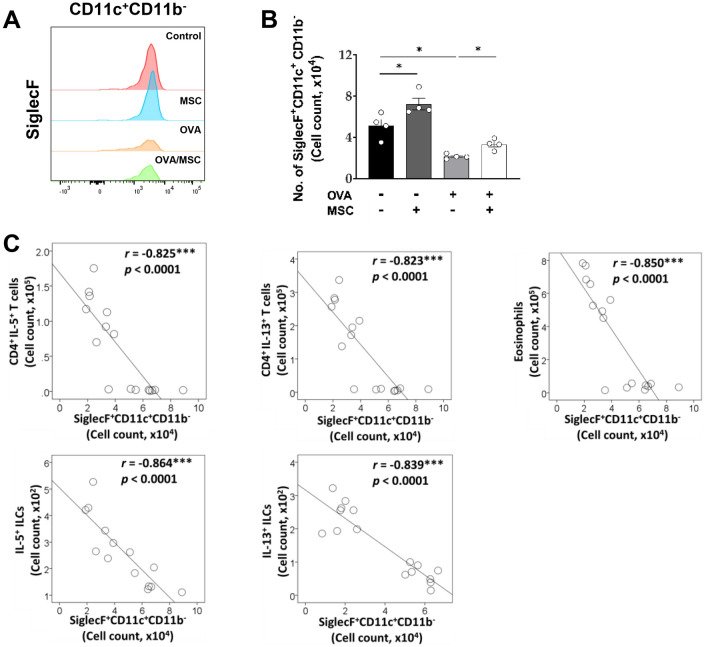
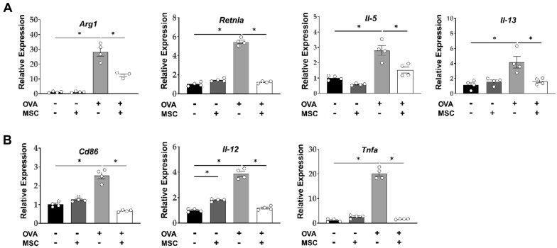
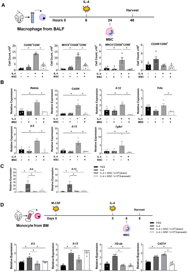
Figure 1Effect of ucMSC on airway hyperresponsiveness and lung inflammation in a murine asthma model. (A) To establish a murine asthma model, 2 mg of Alum and 100 μg of OVA in 100 μL of PBS were injected into the mouse in the peritoneum on days 0 and 7, and 50 μg of OVA in 40 μL of PBS was administered intranasally on days 14, 15, and 16. ucMSCs (105 cells) were injected intratracheally on day 17. (B) Methacholine hyperresponsiveness was measured 24 h after the last intranasal challenge. (C) The number of total inflammatory cells, including macrophages, neutrophils, eosinophils, and lymphocytes in BAL fluid. (D) H&E stain (× 100) of lung histology after allergen challenge. (E) Lung inflammation score in murine asthma model. (i: PBS, ii: ucMSC, iii: OVA, iv: OVA + ucMSC group). n = 4–5 for each group, * indicates P < 0.05, ** indicates P < 0.01. All results are representative of at least three independent experiments. OVA, ovalbumin; Alum, aluminum hydroxide; IP, intraperitoneal; IN, intranasal; MSC, mesenchymal stem cell, RL, resistance of lung; BAL, bronchoavleolar lavage.Figure 2Effect of ucMSCs on Th2 cells and ILC2s by ucMSC treatment in a murine asthma model. (A) The number of lung CD4+ T cells, IL-5+CD4+ T cells, IL-13+CD4+ T cells, IFN-γ+ CD4+ T cells, IL-17A+CD4+ T cells and Foxp3+CD4+ T cells. (B) The number of lung ILCs, IL-5+ ILCs, IL-13+ ILCs, IFN-γ+ ILCs, and IL-17A+ ILCs. (C) ILC2s and ucMSCs co-culture experiment protocol and ILC2 activation, surface marker and Il-10 expression. n = 4–5 for each group, * indicates P < 0.05, ** indicates P < 0.01. All results are representative of at least three independent experiments. MSC, mesenchymal stem cell; ILC, innate lymphoid cell; IL, interleukin; IFN, interferon.Figure 3Effect of ucMSCs on DCs and macrophages by ucMSC treatment in a murine asthma model. (A) The number of lung DC, MHCII+CD86+ DC. (B) AM, MHCII+CD86+ AM. (C) The number of lung M1, M2, and DN and the ratio of M2/M1. (D) The number of M2a, M2b, and M2c macrophages. (E) Correlation plots between M2a macrophages and CD4+IL-5+ T cells, IL-5+ILCs, and eosinophils. (F) Correlation plots between M2c macrophages and CD4+ IL-5+ T cells, IL-5+ILCs, and eosinophils. n = 4–5 for each group, * indicates P < 0.05, ** indicates P < 0.01, *** indicates P < 0.0001. All results are representative of at least three independent experiments. MSC, mesenchymal stem cell; DC, dendritic cells; AM, alveolar macrophage; IL, interleukin; ILC, innate lymphoid cell.Figure 4Effect of ucMSCs on CD11c+CD11b- macrophages in a murine asthma model. (A) Expression level and MFI of SiglecF on CD11c+CD11b- macrophages. (B) The number of SiglecF+CD11c+CD11b- macrophages. (C) Correlation plots between SiglecF+CD11c+CD11b- macrophages and CD4+IL-5+ T cells, CD4+IL-13+ T cells, eosinophils, IL-5+ ILCs, and IL-13+ ILCs. n = 4, * indicates P < 0.05, *** indicates P < 0.0001. All results are representative of at least three independent experiments. MSC, mesenchymal stem cell; IL, interleukin; ILC, innate lymphoid cell.Figure 5Effect of ucMSCs on mRNA expression of lung in a murine asthma model. (A, B) Changes in lung macrophage mRNA expression of (A) Arg1, Retnlα, IL-5, Il-13. (B) Cd86, Il-12, and Tnfa. n = 4 for each group, * indicates P < 0.05. All results are representative of at least three independent experiments. MSC, mesenchymal stem cell; IL, interleukin; TNF: tumor necrosis factor.Figure 6Effect of ucMSCs on mRNA expression of ex vivo and in vitro macrophages. (A, B) Changes in activation markers in the BAL fluid macrophage ex vivo. (C) Analysis of macrophage activation markers using alveolar macrophage cell lines (D) Macrophage differentiation protocol using bone marrow-derived monocytes and changes in their activation markers. n = 4 for each group, * indicates P < 0.05. All results are representative of at least three independent experiments. BAL, Bronchoalveolar lavage; MSC, mesenchymal stem cell; IL, interleukin; TNF: tumor necrosis factor.